


 |
 |
| J Korean Med Assoc > Volume 50(11); 2007 > Article |
Abstract
Modernized urban life style has changed patterns of parasitic infections in Korea. Parasitic diseases caused by soil-transmitted helminths and water-borne protozoans has significantly decreased, while imported parasitic diseases, zoonosis, and opportunistic infections are being increasingly recognized. Tissue-invading helminthiases also invoked formidable health problems, which had been neglected due to the difficult clinical diagnosis and slow progression. However, the diseases are associated with chronic morbidity and severe mortality. A variety of helminths invade the human tissue. With an exception of few entities (i.e., schistosomiasis, clonorchiasis, and paragonimiasis), most of tissue-invading helminths are associated with larvae/juveniles but not with adults. Larval infections might be more serious, since the larvae may migrate throughout the whole body, after which they lodge in critical foci in the brain, eye, liver, or elsewhere or may grow into large masses exerting space-occupying effects (i.e., cysticercosis, sparganosis, and hydatidosis). When the parasites invade the tissue, IgE levels are modulated by several effector molecules including interleukin (IL)-4, IL-6, interferon-γ and other cytokines secreted by different Th-cell subsets. Immediate-type hypersensitivity is related to huge production of Th2-type cytokines, mast cells, eosinophils, and IgE. These immune interactions elicit cellular responses, culminating in immunophysiological changes, which protect the host by surrounding the invasive parasite with granuloma. However, hyperactivation of the immune system may also be harmful to the host, resulting in immune-mediated diseases. This article briefly reviews the biology, clinical manifestations, diagnosis, and principle of the treatment of the tissue-invading helminthic infections, which are important in Korea.
References
1. Ministry of Health and Welfare, Korea Association of Health Promotion. Prevalence of intestinal parasitic diseases in Korea-The seventh Report 2004;Seoul, Korea: Korea Association of Health Promotion.
2. Kwon NH, Oh MJ, Lee SP, Lee BJ, Choi DC. The prevalence and diagnostic value of toxocariasis in unknown eosinophilia. Ann Hematol 2006;85:233-238.
3. Sohn WM, Kim HM, Chung DI, Yee ST. The first human case of Trichinella spiralis infection in Korea. Korean J Parasitol 2000;38:111-115.
4. Chai JY, Han ET, Shin EH, Park JH, Chu JP, Hirota M, Nakamura-Uchiyama F, Nawa Y. An outbreak of gnathostomiasis among Korean emigrants in Myanmar. Am J Trop Med Hyg 2003;69:67-73.
5. Choe G, Lee HS, Seo JK, Chai JY, Lee SH, Eom KS, Chi JG. Hepatic capillariasis: First case in the Republic of Korea. Am J Trop Med Hyg 1993;48:610-625.
6. Sutherst RW. Global change and human vulnerability to vector-borne diseases. Clin Microbiol Rev 2004;17:136-173.
7. Mostafa MH, Sheweita SA. Relationship between schistosomiasis and bladder cancer. Clin Microbiol Rev 1999;12:97-111.
8. Ross AG, Vickers D, Olds GR, Shah SM, McManus DP. Katayama syndrome. Lancet Infect Dis 2007;7:218-224.
9. Goncalves MM, Barreto MG, Peralta RH, Gargioni C, Goncalves T, Igreja RP, Soares MS, Peralta JM. Immunoassays as an auxiliary tool for the serodiagnosis of Schistosoma mansoni infection in individuals with low intensity of egg elimination. Acta Trop 2006;100:24-30.
10. Koukounari A, Sacko M, Keita AD, Gabrielli AF, Landoure A, Dembele R, Clements AC, Whawell S, Donnelly CA, Fenwick A, Traore M, Webster JP. Assessment of ultrasound morbidity indicators of schistosomiasis in the context of large-scale programs illustrated with experiences from Malian children. Am J Trop Med Hyg 2006;75:1042-1052.
11. Choi BI, Han JK, Hong ST, Lee KH. Clonorchiasis and cholangiocarcinoma: Etiologic relationship and imaging diagnosis. Clin Microbiol Rev 2004;17:540-552.
12. Cho SY, Bae JH, Seo BS. Some aspects of human sparganosis in Korea. Korean J Parasitol 1975;13:60-77.
13. Chang KH, Kim WS, Cho SY, Han MC, Kim CW. Comparative evaluation of brain CT and ELISA in the diagnosis of neurocysticercosis. Am J Neuroradiol 1988;9:125-130.
14. Chang KH, Han MH. MRI of CNS parasitic diseases. J Magn Reson Imaging 1998;8:297-307.
15. Cho SY, Kim SI, Kang SY, Choi DY, Suk JS, Choi KS, Ha YS, Chung CS, Myung HJ. Evaluation of enzyme-linked immunosorbent assay in serological diagnosis of human neurocysticercosis using paired samples of serum and cerebrospinal fluid. Korean J Parasitol 1986;24:25-41.
16. Chung JY, Bahk YY, Huh S, Kang SY, Kong Y, Cho SY. A recombinant 10-kDa protein of Taenia solium metacestodes specific to active neurocysticercosis. J Infect Dis 1999;180:1307-1315.
Figure 1
A 52-year-old woman complained of RUQ discomfort and cough. CBC revealed an eosinophilia of 76%. On ultrasonographic examination of the liver, approximately 0.6 cm sized multiple hypodense nodules were noticed. She showed high serum IgG antibody levels against excretory-secretory antigen of L2 stage Toxocara larvae. [Courtesy of Dr. SP Lee (Gacheon University)]
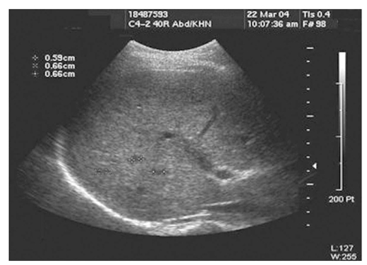
Figure 2
Histopathological finding of Trichinella spiralis muscle larvae. [Courtesy of Dr. WM Sohn (Gyeongsang National University)]
A) Encysted larvae of T. spiralis in the diaphragm of experimental mouse.
B) Pathological examination of the biopsied muscle of a patient infected with T. spiralis showing encysted larvae surrounded by heavily infiltrated inflammatory cells.
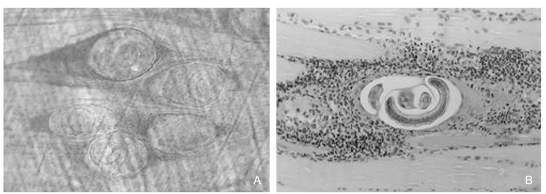
Figure 3
Filarial elephantiasis of the upper and lower extremities. In the final stage of the lymphatic filariasis, tortuous, dilated lymphatic channels resulted in the obstruction of the proximal part. Lymph spaces are obliterated by lymphedema. Fibroblasts might migrate into edematous spaces and caused a thickening of the subcutaneous connective tissue. [Courtesy of Dr. TS Kim (Korea Natonal Institute of Health)]
A) Elephantiasis involving the lower extremities
B) Elephantiasis of the upper extremities
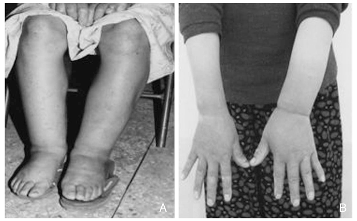
Figure 4
Endoscopic finding of gastric anisakiasis. A long whitish larva is penetrating the gastric mucosal fold in the greater curvature of the mid body. [Courtesy of Dr. WM Sohn (Gyeongsang National University)]

Figure 5
Sonograms of patients with Clonorchis sinensis. [Courtesy of Dr. DI Choi (Sungkyunkwan University) and Dr. ST Hong (Seoul National Univeristy)]
A) Transverse scan of the left hepatic lobe shows moderate dilatation of the intrahepatic bile ducts. Note the slightly hyperechoic bands along dilated ducts, representing increased periductal echogenicity.
B) Oblique scan of the gallbladder shows several floating echogenic foci (arrows), which indicate worms or desquamated materials.
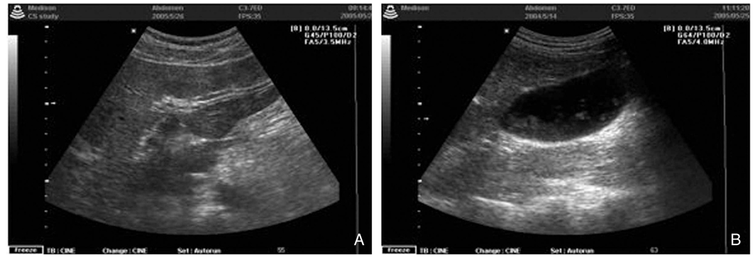
Figure 6
Abdominal CT of a hepatic fascioliasis patient. Huge low density mass suggested a live abscess. Multiple focal tract-like lesions were also observed. The patient revealed high levels of specific anti-Fasciola antibodies in her serum by ELISA.

Figure 7
Pulmonary and cerebral paragonimiasis.
A) Simple chest radiograph shows dense mass-like consolidation, nodules and thin-walled cysts.
B) Consolidations with multiple cysts on chest HRCT.
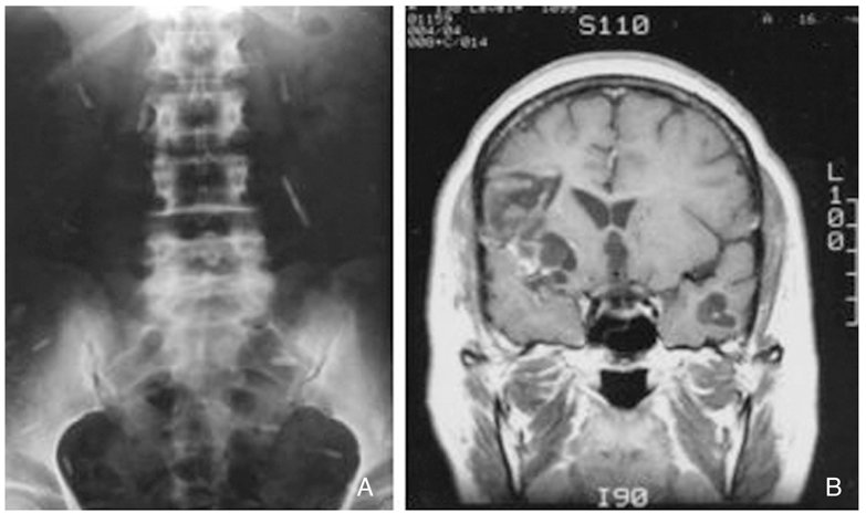
C) Lateral view of simple skull revealed a soup bubble-like calcifications.
D) Calcified granuloma surgically removed from a cerebral case reveals caseous materials filled the granuloma. Smearing of the granuloma wall and content, necrotic parasite eggs are detected.
E) High density conglomerated lesions of calcified granulomas on CT scan.
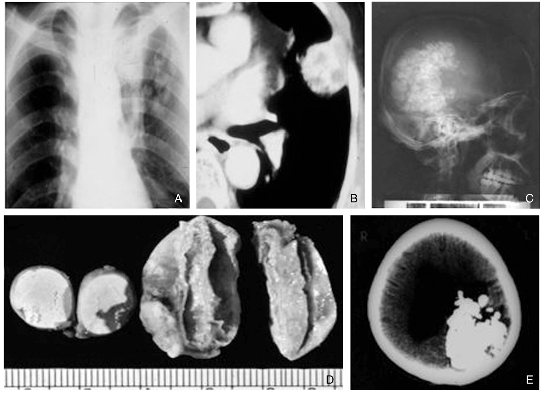
Figure 8
Declining patterns of specific antibody levels in sera of paragonimiasis after treatment against different antigens of P. westermani by ELISA. In most cases of tissue invading helminthic infections, similar serological profiles after the successful treatment could be observed.
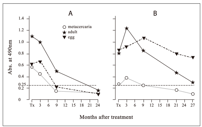
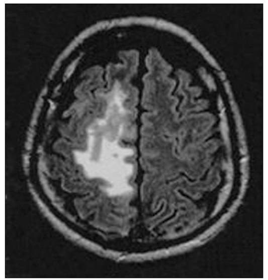
Figure 9
Brain image of neurosparganosis. An extensive area of white matter angioneurotic edema in the left parietal lobe associated with an irregular enhancing mass is observed by brain MRI.
- TOOLS
-
- Share :
-


-
METRICS

-
- 1 Crossref
- Scopus
- 1,071 View
- 3 Download
-

-
Related articles in
J Korean Med Assoc -
Medical Therapy in Thyroid Diseases2000 June;43(6)
Minimally Invasive Surgery in Hepato-Biliary-Pancreatic Disease2003 August;46(8)
Emerging Parasitic Diseases in Korea2007 November;50(11)
Traveling and Imported Parasitic Diseases2007 November;50(11)
Molecular Imaging in Neurodegenerative Diseases2009 February;52(2)
- Editorial Office
-
37 Ichon-ro 46-gil, Yongsan-gu, Seoul
Tel: +82-2-6350-6562 Fax: +82-2-792-5208 E-mail: jkmamaster@gmail.com
Copyright © 2024 by Korean Medical Association.
