


 |
 |
| J Korean Med Assoc > Volume 51(11); 2008 > Article |
Abstract
Basically laboratory vestibular function testing use the vestibular ocular reflex and vestibular spinal reflex like as bedside examination or outpatients' evaluation. Such vestibular laboratory testing can aid diagnosis and can be used to document an abnormality suspected at bedside evaluation. The ability to perform serial vestibular evaluations allows an assessment over time of patients who are undergoing treatment for dizziness or treatment with potentially ototoxic medication. Generally speaking, it includes spontaneous nystagmus, some kinds of evoked nystagmus, ocular eye movement testing, Caloric's testing, rotational chair testing, vestibular evoked myogenic potential, subjective visual vertical, posturography and so on. Those testing have been developed with biomedical engineering based on the proven scientific facts together.
References
1. Fetter Michael. Assessing vestibular function: which tests, when? J Neurol 2000;247:335-342.
2. Kingma Herman. Function tests of the otolith or statolith system. Curr Opin Neurol 2006;19:21-25.
3. Brandt Thomas, Strupp Michael. General vestibular testing. Clinical Neurophysiology 2005;116:406-426.
Figure 1
The results of positional test. It shows two types of horizontal canal benign positional paroxysmal vertigo. The ageotrophic horizontal direction changing positional nystagmus is shown on the top and it means the cupulolithiasis. Otherwise the geotrophic horizontal direction changing positional nystagmus is shown on the bottom and it means the canalithiasis. The lesion sides are right (top) and left (bottom) each.

Figure 2
The results of oculomotor testing. It shows saccadic (top) and pursuit (the middle) tests on left vestibulopathy patient, and then optokinetic (bottom) test on right vestibulopathy patients . Whole graphic data are affected by right beating (top and the middle) and left beating (bottom) spontaneous nystagmus.
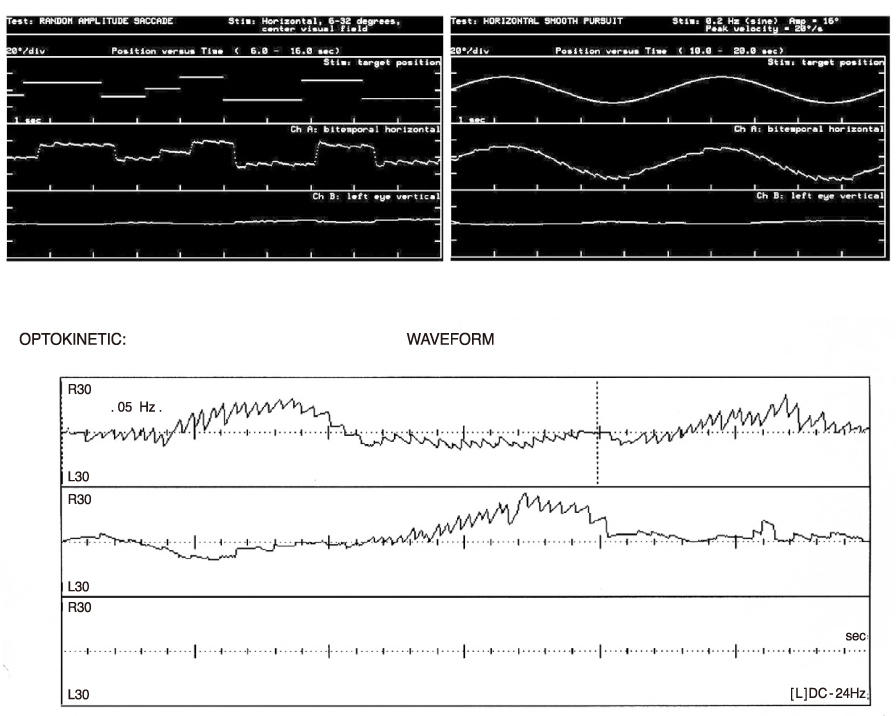
Figure 3
The result of bithermal caloric test. It shows right acute vestibuopathy. The canal paresis and directional preponderance are 78%, 45% each. Visual fixation test is done on left side only and the value is not significant.

Figure 4
The results of sinusoidal harmonic acceleration test. It shows gain (top right), symmetry (top left), phase (bottom) on right acute vestibulopathy patients.

- TOOLS
-
- Share :
-


-
METRICS

-
- 1 Crossref
- Scopus
- 1,066 View
- 0 Download
-

-
Related articles in
J Korean Med Assoc -
Clinical Laboratory Parasitic Examinations1997 February;40(2)
- Editorial Office
-
37 Ichon-ro 46-gil, Yongsan-gu, Seoul
Tel: +82-2-6350-6562 Fax: +82-2-792-5208 E-mail: jkmamaster@gmail.com
Copyright © 2024 by Korean Medical Association.
