


 |
 |
| J Korean Med Assoc > Volume 51(7); 2008 > Article |
Abstract
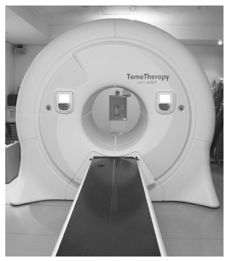
Helical tomotherapy is an image-guided, intensity-modulated radiation therapy delivery system, a hybrid between a linear accelerator and a helical CT scanner. With its unique design features, tomotherapy has improved dose conformity and homogeneity of the target volumes, and conformal avoidance of the adjacent normal tissues. The daily pretreatment megavoltage computed tomography (MVCT) is a powerful tool used in image guided treatment delivery and patient setup verification. If anatomic changes occur during the course of treatment, MVCT images are utilized in correcting target volumes and constructing appropriate adaptive plans. Helical tomotherapy can be applied to more complicated cases, where conventional techniques find their limits: complex tumors with critical organ sparing, simultaneous irradiation of multiple targets, large volume and large superficial tumor irradiation, and recurrent tumor re-irradiation are a few examples. Tomothearpy may change the current paradigm in radiation oncology in the near future. Further studies regarding clinical implementation and treatment outcome of helical tomotherapy will be needed.
References
1. Mackie TR. History of tomotherapy. Phys Med Biol 2006;51:R427-R453.
2. Whitelaw GL, Blasiak-Wal I, Cooke K, Usher C, Macdougall ND, Plowman PN. A dosimetric comparison between two intensity-modulated radiotherapy techniques: tomotherapy vs dynamic linear accelerator. Br J Radiol 2008;81:333-340.
3. Fenwick JD, Tomé WA, Soisson ET, Mehta MP, Mackie TR. Tomotherapy and other innovative IMRT delivery systems. Semin Radiat Oncol 2006;16:199-208.
4. Tomé WA, Jaradat HA, Nelson IA, Ritter MA, Mehta MP. Helical tomotherapy: image guidance and adaptive dose guidance. Front Radiat Ther Oncol 2007;40:162-178.
5. Fenwick JD, Tomé WA, Kissick MW, Mackie TR. Modelling simple helically delivered dose distributions. Phys Med Biol 2005;50:1505-1517.
6. Kron T, Grigorov G, Yu E, Yartsev S, Chen JZ, Wong E, Rodrigues G, Trenka K, Coad T, Bauman G, Van Dyk J. Planning evaluation of radiotherapy for complex lung cancer cases using helical tomotherapy. Phys Med Biol 2004;49:3675-3690.
7. Wieland P, Dobler B, Mai S, Hermann B, Tiefenbacher U, Steil V, Wenz F, Lohr F. IMRT for postoperative treatment of gastric cancer: covering large target volumes in the upper abdomen: a comparison of a step-and-shoot and an arc therapy approach. Int J Radiat Oncol Biol Phys 2004;59:1236-1244.
8. van Vulpen M, Field C, Raaijmakers CP, Parliament MB, Terhaard CH, MacKenzie MA, Scrimger R, Lagendijk JJ, Fallone BG. Comparing step-and-shoot IMRT with dynamic helical tomotherapy IMRT plans for head-and-neck cancer. Int J Radiat Oncol Biol Phys 2005;62:1535-1539.
9. Yartsev S, Kron T, Cozzi L, Fogliata A, Bauman G. Tomotherapy planning of small brain tumours. Radiother Oncol 2005;74:49-52.
10. Soisson ET, Tomé WA, Richards GM, Mehta MP. Comparison of linac based fractionated stereotactic radiotherapy and tomotherapy treatment plans for skull-base tumors. Radiother Oncol 2006;78:313-321.
11. Khoo VS, Oldham M, Adams EJ, Bedford JL, Webb S, Brada M. Comparison of intensity-modulated tomotherapy with stereotactically guided conformal radiotherapy for brain tumors. Int J Radiat Oncol Biol Phys 1999;45:415-425.
12. Fiorino C, Dell'Oca I, Pierelli A, Broggi S, De Martin E, Di Muzio N, Longobardi B, Fazio F, Calandrino R. Significant improvement in normal tissue sparing and target coverage for head and neck cancer by means of helical tomotherapy. Radiother Oncol 2006;78:276-282.
13. Sheng K, Molloy JA, Read PW. Intensity-modulated radiation therapy (IMRT) dosimetry of the head and neck: a comparison of treatment plans using linear accelerator-based IMRT and helical tomotherapy. Int J Radiat Oncol Biol Phys 2006;65:917-923.
14. Lee TK, Rosen II, Gibbons JP, Fields RS, Hogstrom KR. Helical tomotherapy for parotid gland tumors. Int J Radiat Oncol Biol Phys 2008;70:883-891.
15. Mackie TR, Olivera GH, Kapatoes JM. In: Palta JR, Mackie TR, editor. Helical tomotherapy. Intensity-modulated radiation therapy-the state of the art 2003;Madison, WI: Medical Physics Publishing. 247-284.
16. Welsh JS, Bradley K, Ruchala KJ, Mackie TR, Manon R, Patel R, Wiederholt P, Lock M, Hui S, Mehta MP. Megavoltage computed tomography imaging: a potential tool to guide and improve the delivery of thoracic radiation therapy. Clin Lung Cancer 2004;5:303-306.
17. Welsh JS, Lock M, Harari PM, Tomé WA, Fowler J, Mackie TR, Ritter M, Kapatoes J, Forrest L, Chappell R, Paliwal B, Mehta MP. Clinical implementation of adaptive helical tomotherapy: a unique approach to image-guided intensity modulated radiotherapy. Technol Cancer Res Treat 2006;5:465-479.
18. Welsh JS, Patel RR, Ritter MA, Harari PM, Mackie TR, Mehta MP. Helical tomotherapy: an innovative technology and approach to radiation therapy. Technol Cancer Res Treat 2002;1:311-316.
19. Butler EB, Teh BS, Grant WH 3rd, Uhl BM, Kuppersmith RB, Chiu JK, Donovan DT, Woo SY. SMART (simultaneous modulated accelerated radiation therapy) boost: a new accelerated fractionation schedule for the treatment of head and neck cancer with intensity modulated radiotherapy. Int J Radiat Oncol Biol Phys 1999;45:21-32.
20. Chao KS, Ozyigit G, Tran BN, Cengiz M, Dempsey JF, Low DA. Patterns of failure in patients receiving definitive and postoperative IMRT for head-and-neck cancer. Int J Radiat Oncol Biol Phys 2003;55:312-321.
21. Claus F, De Gersem W, De Wagter C, Van Severen R, Vanhoutte I, Duthoy W, Remouchamps V, Van Duyse B, Vakaet L, Lemmerling M, Vermeersch H, De Neve W. An implementation strategy for IMRT of ethmoid sinus cancer with bilateral sparing of the optic pathways. Int J Radiat Oncol Biol Phys 2001;51:318-331.
22. Dawson LA, Anzai Y, Marsh L, Martel MK, Paulino A, Ship JA, Eisbruch A. Patterns of local-regional recurrence following parotid-sparing conformal and segmental intensity-modulated radiotherapy for head and neck cancer. Int J Radiat Oncol Biol Phys 2000;46:1117-1126.
23. Eisbruch A, Marsh LH, Dawson LA, Bradford CR, Teknos TN, Chepeha DB, Worden FP, Urba S, Lin A, Schipper MJ, Wolf GT. Recurrences near base of skull after IMRT for head-and-neck cancer: implications for target delineation in high neck and for parotid gland sparing. Int J Radiat Oncol Biol Phys 2004;59:28-42.
24. Lee N, Xia P, Quivey JM, Sultanem K, Poon I, Akazawa C, Akazawa P, Weinberg V, Fu KK. Intensity-modulated radiotherapy in the treatment of nasopharyngeal carcinoma: an update of the UCSF experience. Int J Radiat Oncol Biol Phys 2002;53:12-22.
25. Schultheiss TE, Wong J, Liu A, Olivera G, Somlo G. Image-guided total marrow and total lymphatic irradiation using helical tomotherapy. Int J Radiat Oncol Biol Phys 2007;67:1259-1267.
26. Wong JY, Liu A, Schultheiss T, Popplewell L, Stein A, Rosenthal J, Essensten M, Forman S, Somlo G. Targeted total marrow irradiation using three-dimensional image-guided tomographic intensity-modulated radiation therapy: an alternative to standard total body irradiation. Biol Blood Marrow Transplant 2006;12:306-315.
27. Smith KS, Gibbons JP, Gerbi BJ, Hogstrom KR. Measurement of superficial dose from a static tomotherapy beam. Medical physics 2008;35:769-774.
28. Ramsey CR, Seibert RM, Robison B, Mitchell M. Helical tomotherapy superficial dose measurements. Medical physics 2007;34:3286-3293.
29. Scrimger RA, Tomé WA, Olivera GH, Reckwerdt PJ, Mehta MP, Fowler JF. Reduction in radiation dose to lung and other normal tissues using helical tomotherapy to treat lung cancer, in comparison to conventional field arrangements. Am J Clin Oncol 2003;26:70-78.
Figure 3
Shifting in parotid gland location due to patient's weight loss after radiation therapy for nasopharyngeal cancer: MVCT merged images (A) before, and (B) during tomotherapy (arrow: parotid gland).
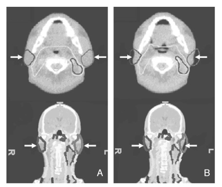
Figure 6
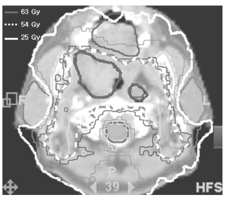
Tomotherapy planning in a complicated case of multiple metastases including sternum and the whole spine.
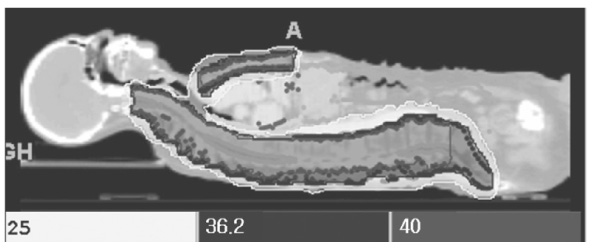
Figure 8
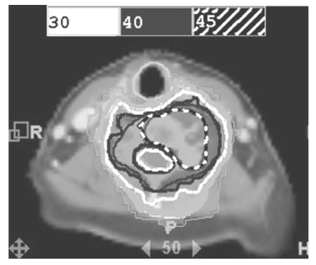
Comparisons of 3D conformal RT, LINAC based IMRT and tomotherapy for whole abdominal irradiation, using specific organ dose-volume histograms (A) Liver (B) Left kidney (C) Bone marrow.

Figure 10
A 72-year-old man who received tomotherapy for scalp angiosarcoma (A) before, (B) immediately after, (C) 3 months after tomotherapy.

- TOOLS
-
- Share :
-


-
METRICS

-
Related articles in
J Korean Med Assoc -
Biological Model and Pharmacotherapy in Internet Addiction2006 March;49(3)
Introduction of intensity modulated radiation therapy2011 November;54(11)
- Editorial Office
-
37 Ichon-ro 46-gil, Yongsan-gu, Seoul
Tel: +82-2-6350-6562 Fax: +82-2-792-5208 E-mail: jkmamaster@gmail.com
Copyright © 2024 by Korean Medical Association.
