


 |
 |
| J Korean Med Assoc > Volume 52(1); 2009 > Article |
Abstract
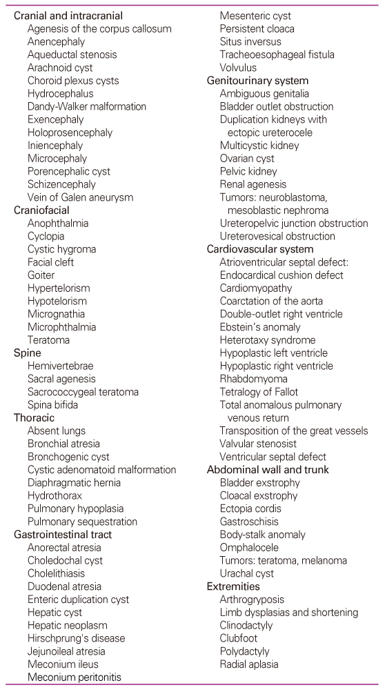
The frequency of fetal malformations accounts for around 3~5% and evaluation of the health of the fetus and screening for fetal malformations has become an important part of prenatal care. Fetal malformations can be classified into structural and chromosomal abnormalities. Improvements in prenatal diagnosis have allowed identification of malformations in fetuses during the first and second trimesters of pregnancy. In prenatal diagnosis, both screening and diagnostic procedures are included. Screening tests include maternal serum aneuploidy screening tests, which are double marker test, triple test, and quadruple test. Recently, first trimester combined ultrasound-biochemical screening and integrated screening were introduced and provided higher detection rates of chromosomal anomalies (ex. Down syndrome). Diagnostic tests are usually performed when screening results are positive and they include chorionic villus sampling, amniocentesis, and percutaneous fetal blood sampling. With high-resolution ultrasound equipment, it is now possible to diagnose most structural abnormalities prenatally. On top of that, recent advances in 3D/4D ultrasound have allowed better understanding of fetal anatomy. However, when ultrasound is equivocal, fetal MRI also can be a useful adjuvant in evaluating fetal structural anomalies. Advances in prenatal diagnostic testing have resulted in tremendous benefits to patients and challenges to healthcare providers and new approaches to education and counseling are needed to assure that all patients receive a complete and balanced review of their prenatal diagnostic testing options.
This article provides an overview of various screening and diagnostic methods for prenatal diagnosis of fetal malformations.
References
1. Korean society of Obstetrics and Gynecology. Obstetrics 2007;4th ed. Seoul: Koonja Publishing. 193-197.
2. Reddy Uma M., Filly Roy A., Copel Joshua A.. Prenatal Imaging : Ultrasonography and Magnetic Resonance Imaging. American College of Obstetricans and Gynecologists. Obstet Gynecol 2008;112:145-157.
3. Ewigman BG, Crane JP, Frigoletto FD, LeFevre ML, Bain RP, McNellis D. Effect of prenatal ultrasound screening on perinatal outcome. N Engl J Med 1993;329:821-827.
4. Diagnostic ultrasound imaging in pregnancy National Institutes of Health Consensus Development Conference Statement February 6-8, 1984 Available at: http://www.ncbi.nlm.nih.gov/books/bv.fcgi?indexed=google&rid=hstat6.chapter.49461. Retrieved February 27, 2007.
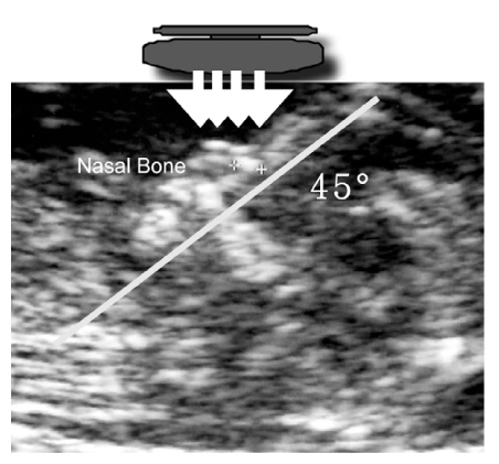
5. Cicero S, Curcio P, Papageorghiou A, Soneck J, Nicolaides K. Absence of nasal bone in fetuses with trisomy 21 at 11~14 weeks of gestation: an observational study. Lancet 2001;358:1665-1667.
6. Cicero S, Spencer K, Avgidou K, Faiola S, Nicolaides K. Maternal serum biochemistry at 11-13+6 weeks in relation to the presence or absence of the fetal nasal bone on ultrasonography in chromosomally abnormal fetuses: an updated analysis of integrated ultrasound and biochemical screening. Prenat Diagn 2005;25:977-983.
7. Pitkin RM. Screening and detection of congenital malformation. Am J Obstet Gynecol 1991;164:1045-1048.
8. Campbell S, Pearce JM. The prenatal diagnosis of fetal structural anomalies by ultrasound. Clin Obstet Gynaecol 1983;10:475-506.
9. Hill LM, Breckle R, Gehrkng WC. Prenatal detection of congenital malformations by ultrasonography. Am J Obstet Gynecol 1985;151:44-50.
10. Manchester DK, Pretorius DH, Avery C, Manco-Johnson ML, Wiggins J, Meier PR, Clewell WH. Accuracy of ultrasound diagnoses in pregnancies complicated by suspected fetal anomalies. Prenat Diagn 1988;8:109-117.
11. Sabbagha RE, Sheikh Z, Tamura RK. Predictive value, sensitivity, and specificity of ultrasonic targeted imaging for fetal anomalies in gravid women at high risk for birth defects. Am J Obstet Gynecol 1985;152:822-827.
12. Filly RA, Cardorza JD, Goldstein RB, Barkovich AJ. Detection of fetal central nervous system anomalies: A practical level of effort for a routine sonogram. Radiology 1989;172:309-311.
13. Yeo L, Vintzileos AM. The use of genetic sonography to reduce the need for amniocentesis in women at high-risk for Down syndrome. Semin perinatol 2003;27:152-159.
14. Vintzileos AM, Egan JF. Adjusting the risk for trisomy 21 on the basis of second-trimester ultrasonography. Am J Obstet Gynecol 1995;17:1639-1640.
15. Benacerraf BR, Nadel AS, Bromley B. Identification of second trimester fetuses with autosomal trisomy by use of a sonographic scoring index. Radiology 1994;193:135-140.
16. Nyberg DA, Luthy DA, Resta RG, Nyberg BC, Williams MA. Age-adjusted ultrasound risk assessment for fetal Down's syndrome during the second trimester: Description of the method and analysis of 142 cases. Ultrasound Obstet Gynecol 1998;12:8-14.
17. Wax JR, Guilbert J, Mather J, Chen C, Royer D, Steinfeld JD, Ingardia CJ. Efficacy of community-based second trimester genetic ultrasonography in detecting the chromosomally abnormal fetus. J Ultrasound med 2000;19:689-694.
18. Gonçalves LF, Lee W, Espinoza J, Romero R. Three- and 4-dimensional ultrasound in obstetric practice: does it help? J Ultrasound Med 2005;24:1599-1624.
19. Gonçalve LF, Nien JK, Espinoza J, Kusanovic JP, Lee W, Swope B, Soto E, Treadwell MC, Romero R. What does 2-dimensional imaging add to 3-and 4-dimensional obstetric ultrasonography? J Ultrasound Med 2006;25:691-699.
20. Levine D, Barens PD, Madsen JR, Li W, Edelman RR. Fetal central nervous system anomalies: MR imaging augments sonographic diagnosis. Radiology 1997;204:635-642.
21. Sponcer K, Spencer CE, Power M, Dawson C, Nicoladies KH. Screening for chromosomal abnormalities in the first trimester using ultrasound and maternal serum bioche-mistry in a one-step clinic: a review three years prospec-tive experience. BJOG 2003;110:281-286.
22. Van Lith JM, Pratt JJ, Beekhuis JR, Mantingh A. Second-trimester maternal serum immunoreactive inhibin as a marker for fetal Down's syndrome. Prenat Diagn 1992;12:801-806.
23. Wald NJ, Densem JW, George L, Muttukrishna S, Knight PG. Prenatal screening for Down's syndrome using inhibin-A as a serum marker. Prenat Diagn 1996;16:143-153.
24. Malone Fergal D, Wald Nicholas J, Canick Jacob A, Ball Robert H, Nybergl David A. First- and second-trimester evaluation of risk (faster): trial principal results of the NICHD multicenter Down syndrome screening study. Am J of Obstet Gynecol 2003;189:S. 79.
25. Biggio JR, Morris TC, Owen J, Stringer JSA. An outcome analysis of five prenatal screening strategies for trisomy 21 in women younger than 35 years. Am J of Obstet Gynecol 2004;190:721-729.
- TOOLS
-
- Share :
-


-
METRICS

-
- 0 Crossref
- Scopus
- 1,296 View
- 3 Download
-

-
Related articles in
J Korean Med Assoc -
Recent trends in the management of anterior knee pain2023 August;66(8)
Recent Trend in Treatment of Atopic Dermatitis1997 March;40(3)
Recent Trend in Treatment of Allergic Rhinitis1997 March;40(3)
Cytogenetics for Prenatal Diagnosis1999 June;42(6)
Differential Diagnosis of Lymphadenopathy2000 October;43(10)
- Editorial Office
-
37 Ichon-ro 46-gil, Yongsan-gu, Seoul
Tel: +82-2-6350-6562 Fax: +82-2-792-5208 E-mail: jkmamaster@gmail.com
Copyright © 2024 by Korean Medical Association.
