


 |
 |
| J Korean Med Assoc > Volume 52(2); 2009 > Article |
Abstract
The study of pain has recently received much attention, especially in understanding its neurophysiology by using new brain imaging techniques, such as positron emission tomography (PET) and functional magnetic resonance imaging (fMRI), both of which allow us to visualize brain function in vivo. Also the new brain imaging devices allow us to evaluate the patient's pain status and plan to treat patients objectively. Functional activation of brain regions are thought to be reflected by increases in the regional cerebral blood flow in the brain imaging studies. Regional cerebral blood flow increases to noxious stimuli are observed in second somatic (SII) and insular regions and in the anterior cingulate cortex and with slightly less consistency in the first somatic area (S1), motor area, supplementary motor area, prefrontal area, amygdala and contralateral thalamus. These data suggest that pain has multidimensions such as sensory-discrimitive, motivational-affective and cognitive-evaluative.
References
1. Apkarian AV, Shi T. In: Ayrapetian A, Apkarian AV, editor. Thalamocortical connections of the cingulate and insula in relation to nociceptive inputs to the cortex. Pain Mechanisms and Management, Series 1997;Amsterdam: IOS Press. 212-220.
2. Bush G, Luu P, Posner MI. Cognitive and emotional influences in anterior cingulate cortex. Trends Cogn Sci 2000;4:215-222.
3. Wall PD, Melzack R. In: Wall PD, Melzack R, editor. Introduction to Pain. Textbook of pain, 4th ed Series 1999;Edinburgh: Churchill Livingstone. 1-8.
4. Heeger DJ, Ress D. What does fMRI tell us about neuronal activity? Nat Rev Neurosci 2002;3:142-151.
5. Jones AK, Brown WD, Friston KJ, Qi LY, Frackowiak RS. Cortical and subcortical localization of response to pain in man using positron emission tomography. Proc. R. Soc. Lond., Ser. B: Biol Sci 1991;244:39-44.
6. Josephs O, Turner R, Friston K. Event-related fMRI. Hum Brain Mapp 1997;5:243-248.4.
7. Bantick SJ, Wise RG, Ploghaus A, Clare S, Smith SM, Tracey I. Imaging how attention modulates pain in humans using functional MRI. Brain 2002;125:310-319.
8. Becerra LR, Breiter HC, Stojanovic M, Fishman S, Edwards A, Comite AR, Gonzalez RG, Borsook D. Human brain activation under controlled thermal stimulation and habituation to noxious heat: an fMRI study. Magn Reson Med 1999;41:1044-1057.
9. Casey KL, Minoshima S, Berger KL, Koeppe RA, Morrow TJ, Frey KA. Positron emission tomographic analysis of cerebral structures activated specifically by repetitive noxious heat stimuli. J Neurophysiol 1994;71:802-807.
10. Cho ZH, Chung SC, Jones JP, Park JB, Park HJ, Lee HJ, Wong EK, Min BI. New findings of the correlation between acupoints and corresponding brain cortices using functional MRI. Proc Natl Acad Sci USA 1998;95:2670-2673.
11. Cho ZH, Chung SC, Lim DW, Wong EK. Effects of the acoustic noise of the gradient systems on fMRI-a study on auditory, motor, and visual cortices. Magn Reson Med 1998;39:331-335.
12. Bonvento Gilles, Sibson Nicola, Pellerin Luc. Does glutamate image your thought? Trends in neurosciences 2002;25:359-364.
13. Magistretti PJ, Pellerin L. Cellular mechanisms of brain energy metabolism and their relevance to functional brain imaging. Philos Trans R Soc London B Biol. Sci 1999;354:1155-1163.
14. Melzack R, Wall PD. Pain mechanisms: a new theory. Science 1965;150:971-979.
15. Cho ZH, Son YD, Kang CK, Han JY, Wong EK, Kim KH, Yim YK, Bai SJ, Lee UJ, Sung KK, Kim KW. Pain dynamics observed by fMRI: differential regression analysis technique. J Magn Reson Imaging 2003;18:273-283.
16. Petrovic P, Kalso E, Petersson KM, Ingvar M. Placebo and opioid analgesia-Imaging a shared neuronal network. Science 2002;295:1737-1740.
17. Posner MI, Raichle ME. The neuroimaging of human brain function. Proc Natl Acad Sci 1998;95:763-764.
18. Posner MI, Rothbart MK. Attention, self-regulation and consciousness. Proc R Soc Lond, Ser. B: Biol Sci 1998;353:1915-1927.
19. Zubieta JK, Smith YR, Bueller JA, Xu Y, Kilbourn MR, Jewett DM, Meyer CR, Koeppe RA, Stohler CS. Regional mu opioid receptor regulation of sensory and affective dimensions of pain. Science 2001;293:311-315.
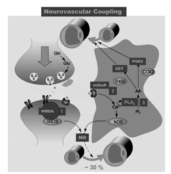
Figure 1
Putative mechanisms involved in neurometabolic and neurovascular coupling during neuronal activation.
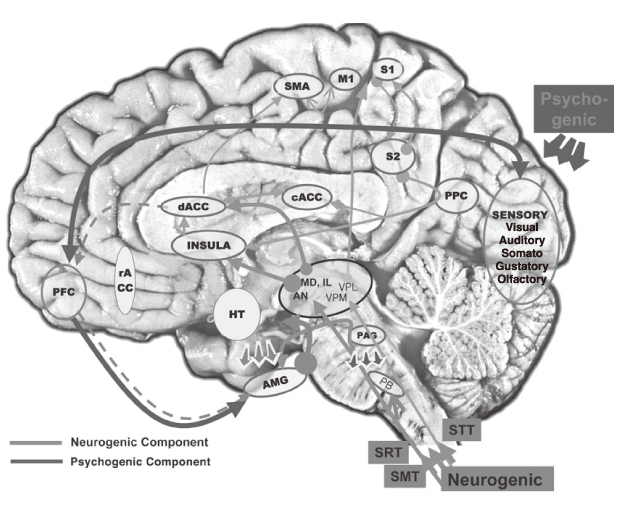
Figure 6
Putative pain pathway in the brain.
PFC: Prefrontal cortex, AMG: Amygdala, rACC: rostral Anterior cyngulate gyrus, dACC: dorsal Anterior cingulate gyrus, cACC: caudal Anterior cingulate gyrus, S1: Sensory area 1, S2: Sensory area 2, SMA: Supplementary motor area, HT: Hypothalamus, STT: Spinothalamic tract, SRT: Spinoreticular tract, SMT: Spinomesencephalic tract.
- TOOLS
-
- Share :
-


-
METRICS

-
- 0 Crossref
- Scopus
- 1,039 View
- 1 Download
-

-
Related articles in
J Korean Med Assoc -
New Trends of Treatments in Sleep Disorder Breathing2000 June;43(6)
New Trends in Breast Imaging2003 June;46(6)
The Proper Use of PET/CT in Tumoring Imaging2004 September;47(9)
- Editorial Office
-
37 Ichon-ro 46-gil, Yongsan-gu, Seoul
Tel: +82-2-6350-6562 Fax: +82-2-792-5208 E-mail: jkmamaster@gmail.com
Copyright © 2024 by Korean Medical Association.
