


 |
 |
| J Korean Med Assoc > Volume 61(4); 2018 > Article |
Min Joo Kim, MD1 , Ka Hee Yi, MD2
, Ka Hee Yi, MD2
Abstract
As the detection of thyroid nodules increases, it is important to differentiate whether thyroid nodules are malignant or not. Ultrasonography-guided fine-needle aspiration cytology is the standard method to diagnose thyroid nodules. Ultrasonographic findings of thyroid nodules can predict the risk of malignancy, and fine-needle aspiration allows the examination of cytopathology of thyroid nodules. However, both are not perfect, with a certain degree of false negative or false positive results. Therefore, we can face thyroid nodules with discordant results of ultrasonographic and fine-needle aspiration findings. In the case of benign features on ultrasonography with malignant cytology, follicular thyroid cancer, follicular variant papillary thyroid cancer, cystic or degenerative changes of thyroid cancer, and thyroiditis are candidates for diagnosis. In contrast, for the nodules with ultrasonographic features of highly suspicious of malignancy but benign cytology, we can consider the possibility of thyroiditis, changes of benign nodule, and cystic changes of thyroid cancer. These various conditions may result in discordant results of ultrasonographic features and fine-needle aspiration cytology, which need special attention not to miss the diagnosis of malignant nodules.
References
1. Ahn HS, Kim HJ, Welch HG. Korea's thyroid-cancer “epidemic”: screening and overdiagnosis. N Engl J Med 2014;371:1765-1767.


2. Shin JH, Baek JH, Chung J, Ha EJ, Kim JH, Lee YH, Lim HK, Moon WJ, Na DG, Park JS, Choi YJ, Hahn SY, Jeon SJ, Jung SL, Kim DW, Kim EK, Kwak JY, Lee CY, Lee HJ, Lee JH, Lee JH, Lee KH, Park SW, Sung JY. Korean Society of Thyroid Radiology (KSThR) and Korean Society of Radiology. Ultrasonography diagnosis and imaging-based management of thyroid nodules: revised Korean Society of thyroid radio-logy consensus statement and recommendations. Korean J Radiol 2016;17:370-395.


3. Cibas ES, Ali SZ. The 2017 Bethesda system for reporting thyroid cytopathology. Thyroid 2017;27:1341-1346.
4. Lee YJ, Kim DW, Park YM, Park HK, Jung SJ, Kim DH, Lee SM, Oh M. Comparison of sonographic and cytological diagnoses of solid thyroid nodules: emphasis on the discordant cases. Diagn Cytopathol 2015;43:953-959.
5. Yi KH, Lee EK, Kang HC, Koh Y, Kim SW, Kim IJ, Na DG, Nam KH, Park SY, Park JW, Bae SK, Baek SK, Baek JH, Lee BJ, Chung KW, Jung YS, Cheon GJ, Kim WB, Chung JH, Rho YS. 2016 Revised Korean Thyroid Association management guidelines for patients with thyroid nodules and thyroid Cancer. Int J Thyroidol 2016;9:59-126.
6. Haugen BR, Alexander EK, Bible KC, Doherty GM, Mandel SJ, Nikiforov YE, Pacini F, Randolph GW, Sawka AM, Schlumberger M, Schuff KG, Sherman SI, Sosa JA, Steward DL, Tuttle RM, Wartofsky L. 2015 American Thyroid Association management guidelines for adult patients with thyroid nodules and differentiated thyroid cancer: the American Thyroid Association guidelines task force on thyroid nodules and differentiated thyroid cancer. Thyroid 2016;26:1-133.
7. Moon WJ, Na DG, Jung SL, Lee JH, Kim HS. Recommendations for ultrasound-based management of thyroid nodules. Seoul: Korean Radiological Society; 2006.
8. Cibas ES, Ali SZ. NCI Thyroid FNA State of the Science Conference. The Bethesda system for reporting thyroid cytopathology. Am J Clin Pathol 2009;132:658-665.
9. Bongiovanni M, Spitale A, Faquin WC, Mazzucchelli L, Baloch ZW. The Bethesda system for reporting thyroid cytopathology: a meta-analysis. Acta Cytol 2012;56:333-339.
10. Hwang SH, Sung JM, Kim EK, Moon HJ, Kwak JY. Imaging-cytology correlation of thyroid nodules with initially benign cytology. Int J Endocrinol 2014;2014:491508.
11. Kwak JY, Koo H, Youk JH, Kim MJ, Moon HJ, Son EJ, Kim EK. Value of US correlation of a thyroid nodule with initially benign cytologic results. Radiology 2010;254:292-300.
12. Kwak JY, Kim EK, Kim MJ, Hong SW, Choi SH, Son EJ, Oh KK, Park CS, Chung WY, Kim KW. The role of ultrasound in thyroid nodules with a cytology reading of “suspicious for papillary thyroid carcinoma". Thyroid 2008;18:517-522.
13. Cho BY, Choi HS, Park YJ, Lim JA, Ahn HY, Lee EK, Kim KW, Yi KH, Chung JK, Youn YK, Cho NH, Park DJ, Koh CS. Changes in the clinicopathological characteristics and out-comes of thyroid cancer in Korea over the past four decades. Thyroid 2013;23:797-804.
14. Choi YJ, Yun JS, Kim DH. Clinical and ultrasound features of cytology diagnosed follicular neoplasm. Endocr J 2009;56:383-389.
15. Lee SH, Baek JS, Lee JY, Lim JA, Cho SY, Lee TH, Ku YH, Kim HI, Kim MJ. Predictive factors of malignancy in thyroid nodules with a cytological diagnosis of follicular neoplasm. Endocr Pathol 2013;24:177-183.
16. Hong AR, Lim JA, Kim TH, Choi HS, Yoo WS, Min HS, Won JK, Lee KE, Jung KC, Park DJ, Park YJ. The Frequency and Clinical Implications of the BRAF(V600E) Mutation in papillary thyroid cancer patients in Korea over the past two decades. Endocrinol Metab (Seoul) 2014;29:505-513.
17. Kim DS, Kim JH, Na DG, Park SH, Kim E, Chang KH, Sohn CH, Choi YH. Sonographic features of follicular variant papillary thyroid carcinomas in comparison with conventional papillary thyroid carcinomas. J Ultrasound Med 2009;28:1685-1692.
18. Yang J, Schnadig V, Logrono R, Wasserman PG. Fine-needle aspiration of thyroid nodules: a study of 4703 patients with histologic and clinical correlations. Cancer 2007;111:306-315.
19. Kapan M, Onder A, Girgin S, Ulger BV, Firat U, Uslukaya O, Oguz A. The reliability of fine-needle aspiration biopsy in terms of malignancy in patients with Hashimoto thyroiditis. Int Surg 2015;100:249-253.
20. Yi KI, Ahn S, Park DY, Lee JC, Lee BJ, Wang SG, Cha W. False-positive cytopathology results for papillary thyroid carcinoma: a trap for thyroid surgeons. Clin Otolaryngol 2017;42:1153-1160.
21. Recavarren RA, Houser PM, Yang J. Potential pitfalls of needle tract effects on repeat thyroid fine-needle aspiration. Cancer Cytopathol 2013;121:155-161.
22. Trimboli P, Nasrollah N, Guidobaldi L, Taccogna S, Cicciarella Modica DD, Amendola S, Romanelli F, Lenzi A, Nigri G, Centanni M, Giovanella L, Valabrega S, Crescenzi A. The use of core needle biopsy as first-line in diagnosis of thyroid nodules reduces false negative and inconclusive data reported by fine-needle aspiration. World J Surg Oncol 2014;12:61.
23. Na DG, Kim DS, Kim SJ, Ryoo JW, Jung SL. Thyroid nodules with isolated macrocalcification: malignancy risk and diagnostic efficacy of fine-needle aspiration and core needle biopsy. Ultrasonography 2016;35:212-219.
24. Ha EJ, Baek JH, Lee JH, Song DE, Kim JK, Shong YK, Hong SJ. Sonographically suspicious thyroid nodules with initially benign cytologic results: the role of a core needle biopsy. Thyroid 2013;23:703-708.
25. Kim SY, Kim EK, Kwak JY, Moon HJ, Yoon JH. What to do with thyroid nodules showing benign cytology and BRAF(V600E) mutation? A study based on clinical and radiologic features using a highly sensitive analytic method Surgery 2015;157:354-361.
26. Nikiforov YE, Ohori NP, Hodak SP, Carty SE, LeBeau SO, Ferris RL, Yip L, Seethala RR, Tublin ME, Stang MT, Coyne C, Johnson JT, Stewart AF, Nikiforova MN. Impact of mutational testing on the diagnosis and management of patients with cytologically indeterminate thyroid nodules: a prospective analysis of 1056 FNA samples. J Clin Endocrinol Metab 2011;96:3390-3397.
27. Jara SM, Bhatnagar R, Guan H, Gocke CD, Ali SZ, Tufano RP. Utility of BRAF mutation detection in fine-needle aspiration biopsy samples read as “suspicious for papillary thyroid carcinoma”. Head Neck 2015;37:1788-1793.
28. Nikiforov YE, Steward DL, Robinson-Smith TM, Haugen BR, Klopper JP, Zhu Z, Fagin JA, Falciglia M, Weber K, Nikiforova MN. Molecular testing for mutations in improving the fine-needle aspiration diagnosis of thyroid nodules. J Clin Endocrinol Metab 2009;94:2092-2098.
29. Nam SJ, Kwak JY, Moon HJ, Yoon JH, Kim EK, Koo JS. Large (≥3cm) thyroid nodules with benign cytology: Can Thyroid Imaging Reporting and Data System (TIRADS) help predict false-negative cytology. PLoS One 2017;12:e0186242.
30. Moon HJ, Kim EK, Kim MJ, Kwak JY. Lymphocytic thyroiditis on fine-needle aspiration biopsy of focal thyroid nodules: approach to management. AJR Am J Roentgenol 2009;193:W345-W349.
31. Hwang S, Shin DY, Kim EK, Yang WI, Byun JW, Lee SJ, Kim G, Im SJ, Lee EJ. Focal lymphocytic thyroiditis nodules share the features of papillary thyroid cancer on ultrasound. Yonsei Med J 2015;56:1338-1344.
32. Chung SR, Baek JH, Park HS, Choi YJ, Sung TY, Song DE, Kim TY, Lee JH. Ultrasound-pathology discordant nodules on core-needle biopsy: malignancy risk and management strategy. Thyroid 2017;27:707-713.
33. Kim DW. Benign lesions that mimic thyroid malignancy on ultrasound. Can Assoc Radiol J 2015;66:79-85.
34. Koo JH, Shin JH, Han BK, Ko EY, Kang SS. Cystic thyroid nodules after aspiration mimicking malignancy: sonographic characteristics. J Ultrasound Med 2010;29:1415-1421.
35. Park NH, Kim DW, Park HJ, Lee EJ, Park JS, Park SI, Bae JM, Lee JH. Thyroid cysts treated with ethanol ablation can mimic malignancy during sonographic follow-up. J Clin Ultrasound 2011;39:441-446.
36. Ha EJ, Baek JH, Lee JH, Lee HY, Song DE, Kim JK, Shong YK, Hong SJ. A focal marked hypoechogenicity within an isoechoic thyroid nodule: is it a focal malignancy or not? Acta Radiol 2015;56:814-819.
37. Hatada T, Okada K, Ishii H, Ichii S, Utsunomiya J. Evaluation of ultrasound-guided fine-needle aspiration biopsy for thyroid nodules. Am J Surg 1998;175:133-136.
38. Ha EJ, Baek JH, Lee JH, Kim JK, Kim JK, Lim HK, Song DE, Sung TY, Kim TY, Kim WB, Shong YK. Core needle biopsy can minimise the non-diagnostic results and need for diagnostic surgery in patients with calcified thyroid nodules. Eur Radiol 2014;24:1403-1409.
39. Hong MJ, Na DG, Baek JH, Sung JY, Kim JH. Cytology-ultrasonography risk-stratification scoring system based on fine-needle aspiration cytology and the Korean-thyroid imaging reporting and data system. Thyroid 2017;27:953-959.
Figure 1
Thyroid nodules with discordant results of ultrasonography and fine-needle aspiration (FNA). (A,B) Thyroid nodule showed low suspicion features (Korean Thyroid Imaging Reporting and Data System 3) in ultrasonography, but FNA cytology (×400) was malignant (Bethesda category VI). The final pathology was follicular variant papillary thyroid carcinoma. (C,D) Thyroid nodule showed intermediate suspicion features (Korean Thyroid Imaging Reporting and Data System 4) in ultrasonography, but FNA cytology (×400) was benign (Bethesda category II).
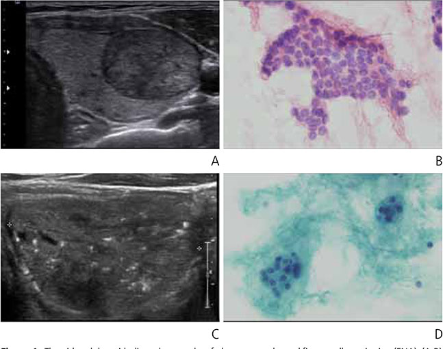
Table 1
Malignant risk of thyroid nodules according to Korean Thyroid Imaging Reporting and Data System
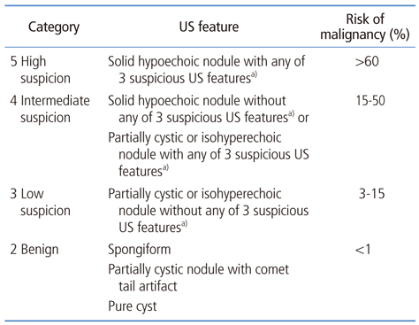
Reproduced from Shin JH, et al. Korean J Radiol 2016;17:370-395, according to the Creative Commons license [2].
US, ultrasonography.
a)Microcalcification, nonparallel orientation (taller-than-wide), spiculated/microlobulated margin.
Table 2
Malignant risk of thyroid nodules according to the Bethesda System Thyroid Cytopathology
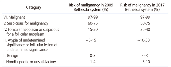
Adapted from Cibas ES, et al. Am J Clin Pathol 2009;132:658-665 [8].
- TOOLS
-
- Share :
-


-
METRICS

-
- 1 Crossref
- Scopus
- 1,948 View
- 28 Download
-

-
Related articles in
J Korean Med Assoc
- Editorial Office
-
37 Ichon-ro 46-gil, Yongsan-gu, Seoul
Tel: +82-2-6350-6562 Fax: +82-2-792-5208 E-mail: jkmamaster@gmail.com
Copyright © 2024 by Korean Medical Association.
