


 |
 |
| J Korean Med Assoc > Volume 47(2); 2004 > Article |
Abstract
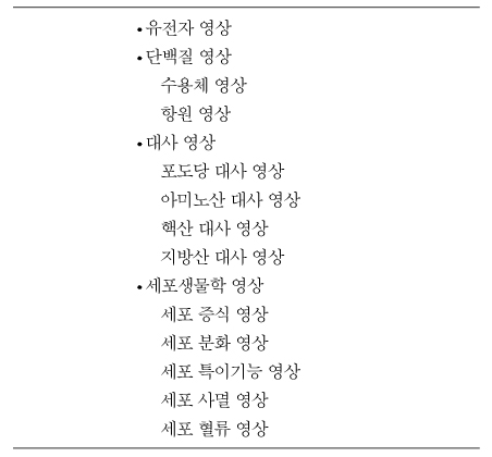
Molecular imaging provides a visualization of normal as well as abnormal cellular processes at a molecular or genetic level rather than at the anatomical level. Molecular imaging is rapidly emerging and a multidisciplinary field coordinating medicine, molecular cell biology, chemistry, pharmacology, genetics, biomedical engineering, and physics. Conventional medical imaging methods utilize the imaging signals produced by nonspecific physico-chemical interaction. However, molecular imaging methods utilize the imaging signals derived from specific cellular or molecular events. Because molecular and genetic changes precede anatomical change in the course of disease development, molecular imaging can detect early events in disease progression. Molecular imaging includes images of proteomics, metabolism, cellular biologic processes as well as genetics. In a narrow sense, molecular imaging means genetic imaging using imaging reporter genes. We can image diverse cellular processes including gene expression, protein-protein interaction, signal transduction pathway, and monitoring of target cell distribution (cancer cells, immune cells, and stem cells) by imaging reporter gene. Molecular imaging methods are classified as optical imaging, nuclear imaging and magnetic resonance imaging. Each imaging modalities have their advantages and weaknesses. In the near future, through molecular imaging we can understand basic mechanisms of disease, and diagnose earlier and, subsequently, treat earlier intractable diseases such as cancer, neuro-degenerative diseases, and immunologic disorders.
References
1. Hadjantonakis AK, Dickinson ME, Fraser SE, Papaioannou VE. Technicolour transgenics: imaging tools for functional genomics in the mouse. Nat Rev Genet 2003;8:613-625.
2. Lok C. Picture perfect. Nature 2001;412:372-374.
3. Gambhir SS, Barrio JR, Phelps ME, Iyer M, Namavari M, Herschman HR, et al. Imaging adenoviral-directed reporter gene expression in living animals with positron emission tomography. Proc Natl Acad Sci USA 1999;96:2333-2338.
4. Tjuvajev JG, Avril N, Oku T, Sasajima T, Miyagawa T, Blasberg RG, et al. Imaging herpes virus thymidine kinase gene transfer and expression by positron emission tomography. Cancer Res 1998;58:4333-4341.
5. Tjuvajev JG, Joshi A, Callegari J, Lindsley L, Joshi R, Blasberg RG, et al. A general approach to the non-invasive imaging of transgenes using cis-linked herpes simplex virus thymidine kinase. Neo-plasia 1999;1:315-312.
6. Yu Y, Annala AJ, Barrio JR, Toyokuni T, Satyamurthy N, Gambhir SS, et al. Quantification of target gene expression by imaging reporter gene expression in living animals. Nat Med 2000;6:933-937.
7. Liang Q, Satyamurthy N, Barrio JR, Toyokuni T, Phelps MP, Herschman HR, et al. Noninvasive, quantitative imaging in living animals of a mutant dopamine D2 receptor reporter gene in which ligand binding is uncoupled from signal transduction. Gene Ther 2001;8:1490-1498.
8. Chung JK. Sodium iodide symporter: Its role in nuclear medicine. J Nucl Med 2002;43:1188-1120.
9. Jacobs A, Voges J, Reszka R, Lercher M, Gossmann A, Heiss WD, et al. Positron-emission tomography of vector-mediated gene expression in gene therapy for gliomas. Lancet 2001;358:727-729.
10. Doubrovin M, Ponomarev V, Beresten T, Balatoni J, Bornmann W, Gelovani Tjuvajev, et al. Imaging transcriptional regulation of p53-dependent genes with positron emission tomography in vivo. Proc Natl Acad Sci USA 2001;98:9300-9305.
11. Kim KI, Chung JK. Evaluation of endogenous p53 expression level using NIS as an imaging reporter gene. SNM 2003;51:88.
12. Ponomarev V, Doubrovin M, Lyddan C, Beresten T, Balatoni J, Tjuvajev JG, et al. Imaging TCR-dependent NFAT-mediated T-cell activation with positron emission tomography in vivo. Neoplasia 2001;3:480-488.
13. Miller MJ, Wei SH, Cahalan MD, Parker I. Autonomous T cell trafficking examined in vivo with intravital two-photon microscopy. Proc Natl Acad Sci USA 2003;100:2604-2609.
14. Miller MJ, Wei SH, Parker I, Cahalan MD. Two-Photon Imaging of Lymphocyte Motility and Antigen Response in Intact Lymph Node. Science 2002;296:1869-1873.
15. Dubey P, Su H, Adonai N, Du S, Rosato A, et al. Quantitative imaging of the T cell antitumor response by positron-emission tomography. Proc Natl Acad Sci USA 2003;100:1232-1237.
16. Sorger JM, McVeigh ER, Hill JM. MRI tracking of mesenchymal stem cell homing to myocardial infarction. SMI 2003;2:221.
17. Paulmurugan R, Umezawa Y, Gambhir SS. Noninvasive imaging of protein-protein interactions in living subjects by using reporter protein complementation and reconstitution strategies. Proc Natl Acad Sci USA 2002;99:15608-15613.
18. Paulmurugan R, Massoud TF, Huang J, Gambhir SS. Modulation of a split synthetic Renilla luciferase complementation-based protein-protein interaction using the small molecule rapamycin in cells and in living animals. SMI 2003;2:198.
- TOOLS
-
- Share :
-


-
METRICS

-
- 0 Crossref
- Scopus
- 1,172 View
- 2 Download
-

-
Related articles in
J Korean Med Assoc -
Perspectives of Biomedical Research2000 January;43(1)
Futrue Perspectives for the Korean Medical Association2000 June;43(6)
Current Issues and Perspectives on School Health Programs2003 February;46(2)
Optical Imaging in the Field of Molecular Imaging2004 February;47(2)
- Editorial Office
-
37 Ichon-ro 46-gil, Yongsan-gu, Seoul
Tel: +82-2-6350-6562 Fax: +82-2-792-5208 E-mail: jkmamaster@gmail.com
Copyright © 2024 by Korean Medical Association.
