


 |
 |
| J Korean Med Assoc > Volume 54(6); 2011 > Article |
Abstract
With the advancement of modern medicine, there have been increasing demands for reconstructive surgeries. The operative technique using free flaps makes it possible for reconstructive surgeons to restore various defects and deformities more precisely. Furthermore, functional problems, such as facial paralysis and lymphedema, can be managed with microsurgical procedures. The need for the composite tissue allograft, including that of the face, has been noticed, and this transplantation surgery required complex microsurgical procedures. With the very high success rate of free flap and popularization of perforator flap, which provides improved outcomes, reconstructive microsurgeons now play major role in various reconstructive fields.
References
1. Chung CN. Clinical observation on skin graft and flap. J Korean Soc Plast Reconstr Surg 1974;1:23-39.
2. Lim P, Cho MJ, Cho HS, Hur E, Lee JW, Ham KS. An axillary scar contracture corrected by microvascular free groin flap transfer. J Korean Soc Plast Reconstr Surg 1978;5:189-194.
3. Park CG, Lee HW, Kim CH. Microvascular free flaps for the reconstruction of head and neck. J Korean Soc Plast Reconstr Surg 1985;12:187-198.
4. Choi SH, Hong SH, Na SK, Park CS. Clinical experiences of dorsalis pedis free flap transfer. J Korean Soc Plast Reconstr Surg 1981;8:189-198.
5. Lee DJ, Hur E, Cho MJ, Lim P. Clinical experiences of free flap transfer. J Korean Soc Plast Reconstr Surg 1980;7:313-320.
6. Franklin JD, Withers EH, Madden JJ Jr, Lynch JB. Use of the free dorsalis pedis flap in head and neck repairs. Plast Reconstr Surg 1979;63:195-204.
7. Leeb DC, Ben-Hur N, Mazzarella L. Reconstruction of the floor of the mouth with a free Dorsalis pedis flap. Plast Reconstr Surg 1977;59:379-381.
8. Ahn HC, Yang EZ, Kim CY, Lee JH. Various applications of deep inferior epigastric artery perforator free flap. J Korean Soc Plast Reconstr Surg 2009;36:707-713.
9. Choi BK, Kim YS, Lee WJ, Lew DH, Tark KC. The Safety of microsurgical head and neck reconstruction in the elderly patients. J Korean Soc Plast Reconstr Surg 2006;33:289-293.
10. Jeon BJ, Lim SY, Hyon WS, Bang SI, Oh KS, Mun GH. Anterolateral thigh flap: our experiences in head and neck reconstruction. J Korean Soc Plast Reconstr Surg 2006;33:276-282.
11. Tark KC, Yoon JS, Shin KS. Analysis of 83 consecutive free flaps. J Korean Soc Plast Reconstr Surg 1993;20:155-166.
12. Koshima I, Soeda S. Inferior epigastric artery skin flaps without rectus abdominis muscle. Br J Plast Surg 1989;42:645-648.
13. Kroll SS, Rosenfield L. Perforator-based flaps for low posterior midline defects. Plast Reconstr Surg 1988;81:561-566.
14. Lee KS, Chang JS, Park JW. Functioning gracilis musculocutaneous free flap transplantation for the reconstruction of injured upper extremity. J Korean Orthop Assoc 1992;27:1868-1876.
15. Jung KM, Hong IP, Kim JH. Reconstruction of extensive lower extermity soft tissue defect using free latissimus dorsi muscle flap with STSG. J Korean Soc Plast Reconstr Surg 1999;26:927-932.
16. Cho KS, Kim DY, Lee SY, Cho BH. Reconstruction of the tissue defects of extremities with anterolateral thigh free flap. J Korean Soc Plast Reconstr Surg 1999;26:281-286.
17. Kim HS, Kim KC, Kim SE. Soft tissue reconstruction of children's extremity with perforator free flap. J Korean Soc Microsurg 2007;16:14-22.
18. Oh CH, Shim JS, Park DH. Lower extremity reconstruction by perforator based flap. J Korean Soc Plast Reconstr Surg 2009;36:720-726.
19. Whitaker IS, Smit JM, Rozen W, Dimopoulou A, Acosta R. Pre operative computed tomographic angiography (CTA): a valuable lesson in planning DIEP flaps. J Plast Reconstr Aesthet Surg 2009;62:551.
20. Lim SY, Pyon JK, Mun GH, Bang SI, Oh KS. Surgical treatment of angiosarcoma of the scalp with superficial parotidectomy. Ann Plast Surg 2010;64:180-182.
21. Kim YH, Kim JT. Perforator flaps in head and neck reconstruction. Hanyang Med Rev 2009;29:265-273.
22. Thorwarth M, Eulzer C, Bader R, Wolf C, Schmidt M, Schultze-Mosgau S. Free flap transfer in cranio-maxillofacial surgery: a review of the current data. Oral Maxillofac Surg 2008;12:113-124.
23. Lipa JE, Butler CE. Enhancing the outcome of free latissimus dorsi muscle flap reconstruction of scalp defects. Head Neck 2004;26:46-53.
24. Lee SH, Mun GH. Transverse thoracodorsal artery perforator flaps: experience with 31 free flaps. J Plast Reconstr Aesthet Surg 2008;61:372-379.
25. Amin A, Rifaat M, Civantos F, Weed D, Abu-Sedira M, Bassiouny M. Free anterolateral thigh flap for reconstruction of major craniofacial defects. J Reconstr Microsurg 2006;22:97-104.
26. Koshima I, Inagawa K, Urushibara K, Moriguchi T. Paraumbilical perforator flap without deep inferior epigastric vessels. Plast Reconstr Surg 1998;102:1052-1057.
27. Mun GH, Lim SY, Hyon WS, Bang SI, Oh KS. A novel reconstruction of 2 distinct defects: concomitant use of a thoracodorsal artery perforator flap and its corresponding muscle flap. Ann Plast Surg 2005;55:676-678.
28. Choi HC, Han SH, Koh KS, Yoon KC, Chung BS. Free muscle flap reconstruction following resection of the skull base tumour. J Korean Soc Plast Reconstr Surg 1993;20:1257-1265.
29. Hanasono MM, Sacks JM, Goel N, Ayad M, Skoracki RJ. The anterolateral thigh free flap for skull base reconstruction. Otolaryngol Head Neck Surg 2009;140:855-860.
30. Zhang B, Li DZ, Xu ZG, Tang PZ. Deep inferior epigastric artery perforator free flaps in head and neck reconstruction. Oral Oncol 2009;45:116-120.
31. Kim KH, Chung CH, Chang YJ, Rho YS. Reconstruction of midfacial defects with free flaps after maxillectomy. J Korean Soc Plast Reconstr Surg 2010;37:607-612.
32. Santamaria E, Granados M, Barrera-Franco JL. Radial forearm free tissue transfer for head and neck reconstruction: versatility and reliability of a single donor site. Microsurgery 2000;20:195-201.
33. Browne JD, Burke AJ. Benefits of routine maxillectomy and orbital reconstruction with the rectus abdominis free flap. Otolaryngol Head Neck Surg 1999;121:203-209.
34. Uglesic V, Virag M, Varga S, Knezevic P, Milenovic A. Reconstruction following radical maxillectomy with flaps supplied by the subscapular artery. J Craniomaxillofac Surg 2000;28:153-160.
35. Suga H, Asato H, Okazaki M, Okochi M, Narushima M. Combination of costal cartilage graft and rib-latissimus dorsi flap: a new strategy for secondary reconstruction of the maxilla. J Craniofac Surg 2007;18:639-642.
36. Andrades P, Rosenthal EL, Carroll WR, Baranano CF, Peters GE. Zygomatic-maxillary buttress reconstruction of midface defects with the osteocutaneous radial forearm free flap. Head Neck 2008;30:1295-1302.
37. Chang DW, Langstein HN. Use of the free fibula flap for restoration of orbital support and midfacial projection following maxillectomy. J Reconstr Microsurg 2003;19:147-152.
38. Maranzano M, Atzei A. The versatility of vascularized iliac crest with internal oblique muscle flap for composite upper maxillary reconstruction. Microsurgery 2007;27:37-42.
39. Pomahac B, Lengele B, Ridgway EB, Matros E, Andrews BT, Cooper JS, Kutz R, Pribaz JJ. Vascular considerations in composite midfacial allotransplantation. Plast Reconstr Surg 2010;125:517-522.
40. Blocker TG Jr, Stout RA. Mandibular reconstruction, World War II. Plast Reconstr Surg (1946) 1949;4:153-156.
41. Shenaq SM, Klebuc MJ. The iliac crest microsurgical free flap in mandibular reconstruction. Clin Plast Surg 1994;21:37-44.
42. Wei FC, Jain V, Celik N, Chen HC, Chuang DC, Lin CH. Have we found an ideal soft-tissue flap? An experience with 672 anterolateral thigh flaps Plast Reconstr Surg 2002;109:2219-2226.
43. Harii K, Ohmori K, Torii S. Free gracilis muscle transplantation, with microneurovascular anastomoses for the treatment of facial paralysis. A preliminary report. Plast Reconstr Surg 1976;57:133-143.
44. Terzis JK. Pectoralis minor: a unique muscle for correction of facial palsy. Plast Reconstr Surg 1989;83:767-776.
45. Harii K, Asato H, Yoshimura K, Sugawara Y, Nakatsuka T, Ueda K. One-stage transfer of the latissimus dorsi muscle for reanimation of a paralyzed face: a new alternative. Plast Reconstr Surg 1998;102:941-951.
46. Choi JH, Minn KW. Chest wall reconstruction with muscle flap. J Korean Soc Plast Reconstr Surg 2003;30:413-419.
47. Sullivan SR, Truxillo TM, Mann GN, Isik FF. Utility of the free deep inferior epigastric perforator flap in chest wall reconstruction. Breast J 2007;13:50-54.
48. Ahn S. Breast reconstruction using implants. J Korean Med Assoc 2011;54:51-60.
49. Cho YK, Yang JD, Kim GR, Chung HY, Cho BC, Park HY. Immediate implant reconstruction using silicone prosthesis in breast cancer patients after skin sparing mastectomy. J Korean Soc Plast Reconstr Surg 2010;37:749-757.
50. Lee HK, Kim YJ, Lee SJ. The latissimus dorsi flap with an implant: the most useful & proper breast reconstruction method for the young mastectomy patient. J Korean Soc Aesthetic Plast Surg 2000;6:83-90.
51. Park JH, Bang SI, Kim SH, Im SY, Mun GH, Hyon WS, Oh KS. Breast reconstruction with the extended latissimus dorsi musculocutancous flap. J Korean Soc Plast Reconstr Surg 2005;32:408-415.
52. Eom JS. Breast reconstruction using pedicled transverse rectus abdominis musculocutaneous (TRAM) flap. J Korean Med Assoc 2011;54:12-21.
53. Kim EK, Eom JS, Ahn SH, Son BH, Lee TJ. Evolution of the pedicled TRAM flap: a prospective study of 500 consecutive cases by a single surgeon in Asian patients. Ann Plast Surg 2009;63:378-382.
54. Nahabedian MY, Momen B, Galdino G, Manson PN. Breast reconstruction with the free TRAM or DIEP flap: patient selection, choice of flap, and outcome. Plast Reconstr Surg 2002;110:466-475.
55. Jin US, Minn KW. Breast reconstruction using the transverse rectus abdominis musculocutaneous (TRAM) free flap. J Korean Med Assoc 2011;54:22-34.
56. Kim JH, Park JU, Cho SH, Eo SR. Immediate breast reconstruction with DIEP free flap. J Korean Soc Microsurg 2008;17:94-100.
57. Gill PS, Hunt JP, Guerra AB, Dellacroce FJ, Sullivan SK, Boraski J, Metzinger SE, Dupin CL, Allen RJ. A 10-year retrospective review of 758 DIEP flaps for breast reconstruction. Plast Reconstr Surg 2004;113:1153-1160.
58. Tajsic N, Winkel R, Hoffmann R, Husum H. Sural perforator flap for reconstructive surgery in the lower leg and the foot: a clinical study of 86 patients with post-traumatic osteomyelitis. J Plast Reconstr Aesthet Surg 2009;62:1701-1708.
59. Wong CH, Tan BK. Perforator-sparing transposition flaps for lower limb defects: anatomic study and clinical application. Ann Plast Surg 2007;58:614-621.
60. Momeni A, Krischak S, Bannasch H. The thoracodorsal artery perforator flap with a vascularized scapular segment for reconstruction of a composite lower extremity defect. Microsurgery 2006;26:515-518.
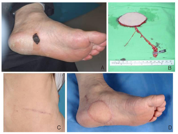
61. Yang D, Yang JF, Morris SF, Tang M, Nie C. Medial plantar artery perforator flap for soft-tissue reconstruction of the heel. Ann Plast Surg 2011;02. 04. [Epub].
62. Unglaub F, Wolf MB, Dragu A, Forst J, Horch RE, Kneser U. Reconstruction of a child's forefoot defect using a distally based pedicled medial plantar flap. Arch Orthop Trauma Surg 2010;130:155-158.
63. Caleffi E, Bocchi A, Montacchini G, Papadia F. Reconstruction of the heel by a medial plantar flap. Ital J Orthop Traumatol 1989;15:191-196.
64. Botte MJ, Gellman H. Reconstruction of a traumatic hallux amputation using a plantar V-Y advancement flap. Clin Orthop Relat Res 1987;220):211-216.
65. Tamura A, Takeuchi Y, Yamakage A. Reconstruction of plantar heel defects with free gracilis musculocutaneous flap. J Foot Ankle Surg 1994;33:274-277.
66. Dubrow TJ, Lesavoy MA. Acne of the heel: acne vulgaris complicating a free vascularized latissimus dorsi musculocutaneous flap. Ann Plast Surg 1989;23:349-351.
67. Ikuta Y, Murakami T, Yoshioka K, Tsuge K. Reconstruction of the heel pad by flexor digitorum brevis musculocutaneous flap transfer. Plast Reconstr Surg 1984;74:86-96.
68. Bostwick J 3rd. Reconstruction of the heel pad by muscle transposition and split skin graft. Surg Gynecol Obstet 1976;143:973-974.
69. Jeong JH. Reconstruction of large heel defects using gracilis muscle free flaps. Yeungnam Univ J Med 1997;14:227-236.
70. Hong JP. Reconstruction of the diabetic foot using the anterolateral thigh perforator flap. Plast Reconstr Surg 2006;117:1599-1608.
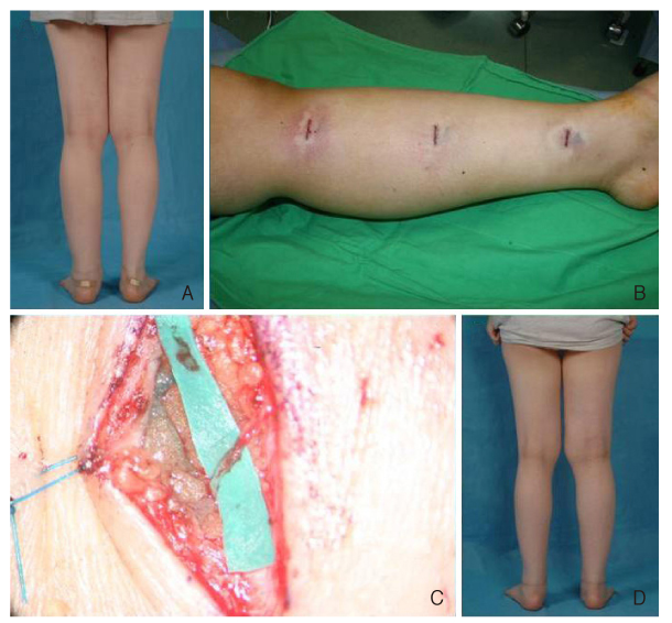
71. Nagase T, Gonda K, Inoue K, Higashino T, Fukuda N, Gorai K, Mihara M, Nakanishi M, Koshima I. Treatment of lymphedema with lymphaticovenular anastomoses. Int J Clin Oncol 2005;10:304-310.
72. Dubernard JM, Owen E, Herzberg G, Martin X, Guigal V, Dawahra M, Pasticier G, Mongin-Long D, Kopp C, Ostapetz A, Lanzetta M, Kapila H, Hakim N. The first transplantation of a hand in humans: early results. Chirurgie 1999;124:358-365.
73. Devauchelle B, Badet L, Lengelé B, Morelon E, Testelin S, Michallet M, D'Hauthuille C, Dubernard JM. First human face allograft: early report. Lancet 2006;368:203-209.
74. Dubernard JM, Lengele B, Morelon E, Testelin S, Badet L, Moure C, Beziat JL, Dakpe S, Kanitakis J, D'Hauthuille C, El Jaafari A, Petruzzo P, Lefrancois N, Taha F, Sirigu A, Di Marco G, Carmi E, Bachmann D, Cremades S, Giraux P, Burloux G, Hequet O, Parquet N, Frances C, Michallet M, Martin X, Devauchelle B. Outcomes 18 months after the firs4t human partial face transplantation. N Engl J Med 2007;357:2451-2460.
75. Soni CV, Barker JH, Pushpakumar SB, Furr LA, Cunningham M, Banis JC Jr, Frank J. Psychosocial considerations in facial transplantation. Burns 2010;36:959-964.
76. Siemionow M, Agaoglu G, Unal S. A cadaver study in preparation for facial allograft transplantation in humans: part II. Mock facial transplantation. Plast Reconstr Surg 2006;117:876-885.
77. Siemionow MZ, Gordon CR. Institutional review board-based recommendations for medical institutions pursuing protocol approval for facial transplantation. Plast Reconstr Surg 2010;126:1232-1239.
Figure 1
Concept change from reconstructive ladder (A) through reconstructive elevator (B) to reconstructive pie.
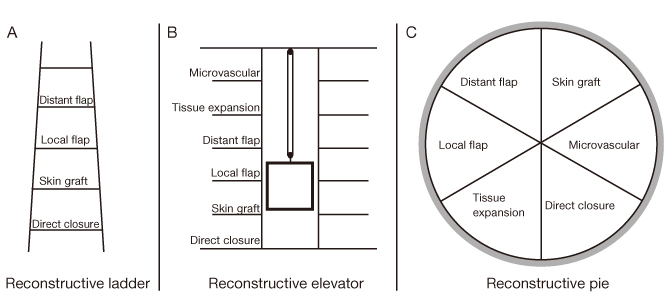
Figure 2
Preoperative planning with computed tomography (CT) angiography. (A) Transverse view of rendered 3D CT angiography of the abdomen. (B) Coronal view of rendered 3D CT angiography. (C) Evaluation of recipient vessels (in this case, internal mammary vessels are used as recipient vessels) can be done with CT angiography. P, perforating vessels.
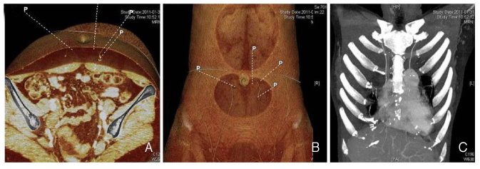
Figure 3
Burn scar contracture, scalp. (A) Preoperative view. (B) Flap design (thoracodorsal artery perforator flap). (C) Elevated flap. (D) Two months postoperative view.
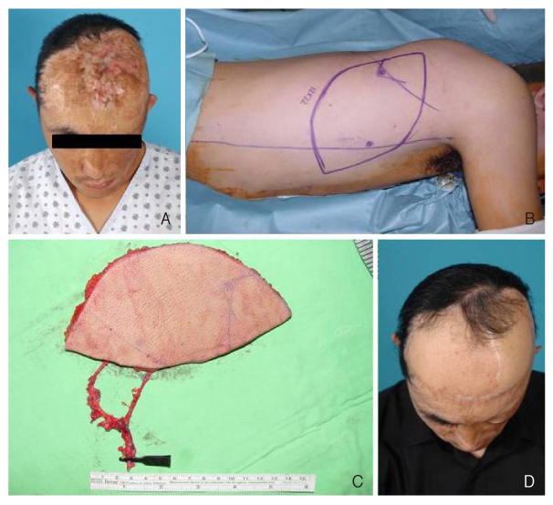
Figure 4
Recurred nasopharyngeal cancer, left temporal region. (A) Defect after subtotal petrosectomy. (B) Chimeric pattern anterolateral thigh flap with vastus lateralis muscle. (C) Elevated muscle flap can be used for obliteration of dead space after ablative surgery. (D) Immediate posto-perative view.
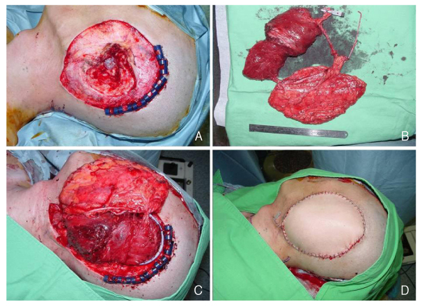
Figure 5
Recurred maxillary cancer, right. (A) Defect after extended radical maxillectomy with orbital exenteration. (B) Elevated vertical rectus abdominis musculocutaneous flap and design. (C,D) Fortyone months postoperative view. (E) Intraoral view.
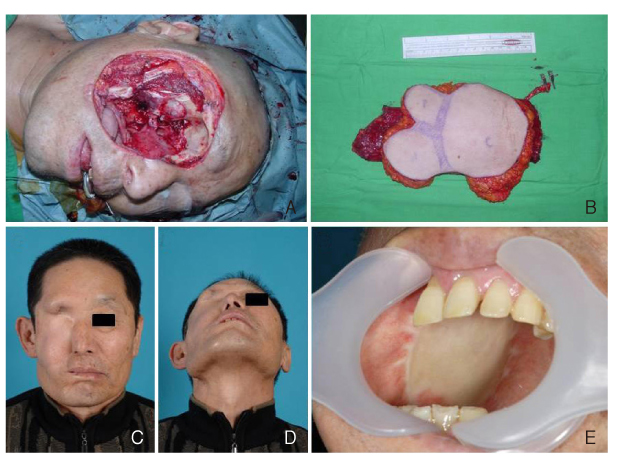
Figure 6
Retromolar trigone cancer, right. (A) Defect after mandibulectomy, right. (B) Flap design, right peroneal region. (C) Elevated fibular osteocutaeous flap. (D) Inset of fibular osteocutaneous flap was done. (E) Three months postoperative view. (F) Split thickness skin graft was done on the donor-site.
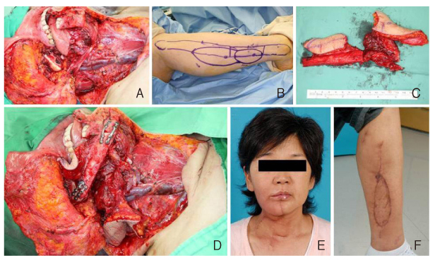
Figure 7
Tongue cancer, right. (A) Defect after near total glossectomy. (B) Preoperative computed tomography angiography was done to select appropriate perforators. (C) Design according to the defect. (D) Elevated anterolateral thigh flap. (E) Inset of the flap. (F) Six weeks postoperative view.
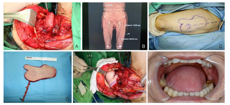
Figure 8
Esophageal cancer. (A) Jejunal free flap can be a successful option for esophageal reronstruction. (B) The esophagogram after reconstruction shows patent luminal structure.
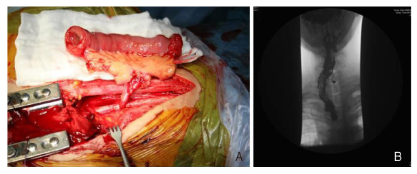
Figure 9
Facial nerve schwannoma, left. (A) Preoperative view. (B) Elevated latissimus dorsi muscle flap with thoracodorsal nerve. (C) Seven months postoperative view.

Figure 10
Fibromatosis, sternal region. (A) Defect after radical excision of mass. (B) Elevated deep inferior epigastric artery perforator flap. (C) Immediate postoperative view.
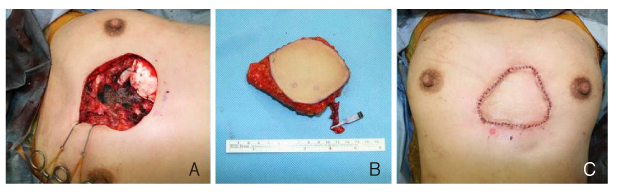
Figure 11
Invasive ductal carcinoma, right. (A) Defect after nipple sparing mastectomy and design of deep inferior epigastric artery perforator flap. (B) Elevated flap. (C,D) Seven months postoperative views.
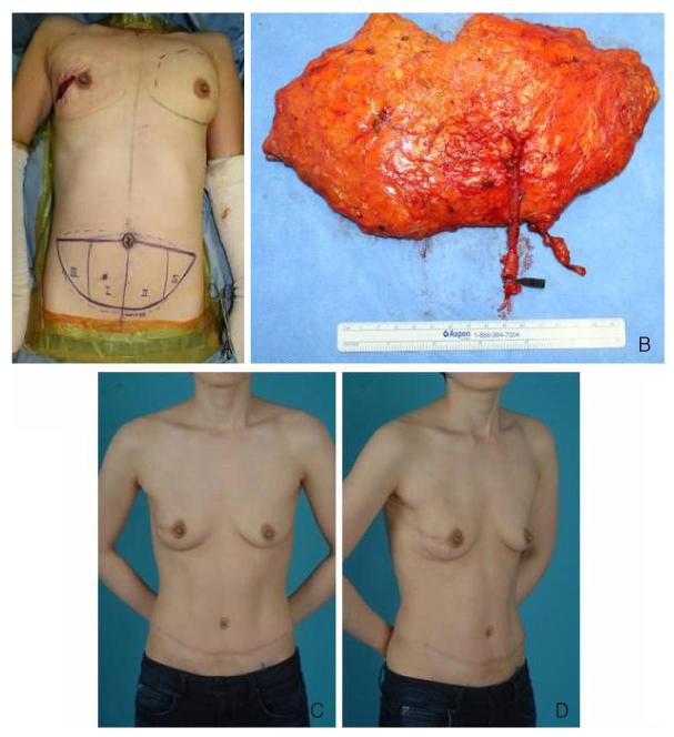
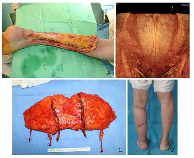
Figure 12
Paraffinoma, left calf. (A) Defect after radical excision. (B) Preoperative rendered CT angiography for selection of appropriate perforators. (C) Elevated deep inferior epigastric perforator flap with 2 deep inferior epigastric vessels and 2 superficial inferior epigastric veins. (D) Two years postoperative view.
- TOOLS
-
- Share :
-


-
METRICS

-
Related articles in
J Korean Med Assoc -
Updated view on the treatment of chronic obstructive pulmonary disease in Korea2021 March;64(3)
Pharmacotherapy of Chronic Obstructive Pulmonary Diseases1998 April;41(4)
The Practice of Alternative Medicine in Korea1998 December;41(12)
Preparation of Physicians for the 21st Century in Korea2000 January;43(1)
Prospects of Medical Suspecialty Board System in Korea2001 November;44(11)
- Editorial Office
-
37 Ichon-ro 46-gil, Yongsan-gu, Seoul
Tel: +82-2-6350-6562 Fax: +82-2-792-5208 E-mail: jkmamaster@gmail.com
Copyright © 2024 by Korean Medical Association.
