


 |
 |
| J Korean Med Assoc > Volume 53(12); 2010 > Article |
Abstract
Musculskeletal magnetic resonance imaging (MRI) applications are making the transition rapidly from 1.5 tesla (T) to 3T. The higher signal-to-noise ratio (SNR) that is available with a 3T MRI system allows for greater spatial resolution and provides the potential to improve the diagnostic capability of musculoskeletal MRI. With the use of 3T systems, one can enhance the SNR, spatial resolution, and contrast-to-noise ratio of intrinsic joint structures such as osseous, tendinous, cartilaginous, and ligamentous structures, which makes them more discernable and amenable to proper radiologic assessment. The SNR gain and coil technology advances allow for a smaller voxel-size and parallel imaging, reducing the acquisition time without significant signal loss. Three-dimensional (3D) fast spin echo sequences with isotropic resolution reduce partial volume artifacts through the acquisition of thin continuous sections and enable free 3D-multiplanar-reformatting without loss of image quality. This technique may be a promising method to replace currently used 2D sequences in clinical practice. In addition to current clinical applications, 3T MRI will contribute to the development of new molecular and functional MRI techniques.
References
1. Mosher TJ. Musculoskeletal imaging at 3T: current techniques and future applications. Magn Reson Imaging Clin N Am 2006;14:63-76.
2. Ramnath RR. 3T MR imaging of the musculoskeletal system (Part II): clinical applications. Magn Reson Imaging Clin N Am 2006;14:41-62.
3. Kladny B, Glückert K, Swoboda B, Beyer W, Weseloh G. Comparison of low-field (0.2 Tesla) and high-field (1.5 Tesla) magnetic resonance imaging of the knee joint. Arch Orthop Trauma Surg 1995;114:281-286.
4. Cotten A, Delfaut E, Demondion X, Lapégue F, Boukhelifa M, Boutry N, Chastanet P, Gougeon F. MR imaging of the knee at 0.2 and 1.5 T: correlation with surgery. AJR Am J Roentgenol 2000;174:1093-1097.
5. Loew R, Kreitner KF, Runkel M, Zoellner J, Thelen M. MR arthrography of the shoulder: comparison of low-field (0.2 T) vs high-field (1.5 T) imaging. Eur Radiol 2000;10:989-996.
6. Shellock FG, Bert JM, Fritts HM, Gundry CR, Easton R, Crues JV 3rd. Evaluation of the rotator cuff and glenoid labrum using a 0.2-Tesla extremity magnetic resonance (MR) system: MR results compared to surgical findings. J Magn Reson Imaging 2001;14:763-770.
7. Kreitner KF, Romaneehsen B, Krummenauer F, Oberholzer K, Müller LP, Düber C. Fast magnetic resonance imaging of the knee using a parallel acquisition technique (mSENSE): a prospective performance evaluation. Eur Radiol 2006;16:1659-1666.
8. Zuo J, Li X, Banerjee S, Han E, Majumdar S. Parallel imaging of knee cartilage at 3 Tesla. J Magn Reson Imaging 2007;26:1001-1009.
9. Lichy MP, Wietek BM, Mugler JP 3rd, Horger W, Menzel MI, Anastasiadis A, Siegmann K, Niemeyer T, Königsrainer A, Kiefer B, Schick F, Claussen CD, Schlemmer HP. Magnetic resonance imaging of the body trunk using a single-slab, 3-dimensional, T2-weighted turbo-spin-echo sequence with high sampling efficiency (SPACE) for high spatial resolution ima-ging: initial clinical experiences. Invest Radiol 2005;40:754-760.
10. Norris DG. High field human imaging. J Magn Reson Imaging 2003;18:519-529.
11. Fullerton GD, Cameron IL, Ord VA. Orientation of tendons in the magnetic field and its effect on T2 relaxation times. Radiology 1985;155:433-435.
12. Kijowski R, Davis KW, Woods MA, Lindstrom MJ, De Smet AA, Gold GE, Busse RF. Knee joint: comprehensive assessment with 3D isotropic resolution fast spin-echo MR imaging: diagnostic performance compared with that of conventional MR imaging at 3.0 T. Radiology 2009;252:486-495.
13. Magee T, Williams D. 3.0-T MRI of meniscal tears. AJR Am J Roentgenol 2006;187:371-375.
14. Ramnath RR, Magee T, Wasudev N, Murrah R. Accuracy of 3-T MRI using fast spin-echo technique to detect meniscal tears of the knee. AJR Am J Roentgenol 2006;187:221-225.
15. Brody JM, Hulstyn MJ, Fleming BC, Tung GA. The meniscal roots: gross anatomic correlation with 3-T MRI findings. AJR Am J Roentgenol 2007;188:W446-W450.
16. Ha TP, Li KC, Beaulieu CF, Bergman G, Ch'en IY, Eller DJ, Cheung LP, Herfkens RJ. Anterior cruciate ligament injury: fast spin-echo MR imaging with arthroscopic correlation in 217 examinations. AJR Am J Roentgenol 1998;170:1215-1219.
17. Vellet AD, Lee DH, Munk PL, Hewett L, Eliasziw M, Dunlavy S, Vidito L, Fowler PJ, Miniaci A, Amendola A. Anterior cruciate ligament tear: prospective evaluation of diagnostic accuracy of middle- and high-field-strength MR imaging at 1.5 and 0.5 T. Radiology 1995;197:826-830.
18. Barry KP, Mesgarzadeh M, Triolo J, Moyer R, Tehranzadeh J, Bonakdarpour A. Accuracy of MRI patterns in evaluating anterior cruciate ligament tears. Skeletal Radiol 1996;25:365-370.
19. Umans H, Wimpfheimer O, Haramati N, Applbaum YH, Adler M, Bosco J. Diagnosis of partial tears of the anterior cruciate ligament of the knee: value of MR imaging. AJR Am J Roentgenol 1995;165:893-897.
20. Servant CT, Ramos JP, Thomas NP. The accuracy of magnetic resonance imaging in diagnosing chronic posterior cruciate ligament injury. Knee 2004;11:265-270.
21. Vahey TN, Broome DR, Kayes KJ, Shelbourne KD. Acute and chronic tears of the anterior cruciate ligament: differential features at MR imaging. Radiology 1991;181:251-253.
22. Gross ML, Grover JS, Bassett LW, Seeger LL, Finerman GA. Magnetic resonance imaging of the posterior cruciate ligament: clinical use to improve diagnostic accuracy. Am J Sports Med 1992;20:732-737.
23. Suh JS, Cho JH, Shin KH, Kim SJ. Chondromalacia of the knee: evaluation with a fat-suppression three-dimensional SPGR imaging after intravenous contrast injection. J Magn Reson Imaging 1996;6:884-888.
24. Gold GE, Han E, Stainsby J, Wright G, Brittain J, Beaulieu C. Musculoskeletal MRI at 3.0 T: relaxation times and image contrast. AJR Am J Roentgenol 2004;183:343-351.
25. Schröder RJ, Fischbach F, Unterhauser FN, Weiler A, Felix R, Bruhn H. Value of various MR sequences using 1.5 and 3.0 Tesla in analyzing cartilaginous defects of the patella in an animal model. Rofo 2004;176:1667-1675.
26. Kijowski R, Blankenbaker DG, Davis KW, Shinki K, Kaplan LD, De Smet AA. Comparison of 1.5-and 3.0-T MR imaging for evaluating the articular cartilage of the knee joint. Radiology 2009;250:839-848.
27. Tardieu M, Lazennec JY, Christel P, Brasseur JL, Roger B, Grenier P. Normal and pathological MRI aspects of the posterolateral corner of the knee. J Radiol 1995;76:605-609.
28. Sims WF, Jacobson KE. The posteromedial corner of the knee: medial-sided injury patterns revisited. Am J Sports Med 2004;32:337-345.
29. Newberg AH, Newman JS. Imaging the painful hip. Clin Orthop Relat Res 2003;406):19-28.
30. Mintz DN, Hooper T, Connell D, Buly R, Padgett DE, Potter HG. Magnetic resonance imaging of the hip: detection of labral and chondral abnormalities using noncontrast imaging. Arthroscopy 2005;21:385-393.
31. Hodler J, Trudell D, Pathria MN, Resnick D. Width of the articular cartilage of the hip: quantification by using fatsuppression spin-echo MR imaging in cadavers. AJR Am J Roentgenol 1992;159:351-355.
32. Rubin SJ, Totterman SM, Meyers SP, Hartley DF. Magnetic resonance imaging of the hip with a pelvic phased-array surface coil: a technical note. Skeletal Radiol 1998;27:77-82.
33. Keeney JA, Peelle MW, Jackson J, Rubin D, Maloney WJ, Clohisy JC. Magnetic resonance arthrography versus arthroscopy in the evaluation of articular hip pathology. Clin Orthop Relat Res 2004;429):163-169.
34. Leunig M, Podeszwa D, Beck M, Werlen S, Ganz R. Magnetic resonance arthrography of labral disorders in hips with dysplasia and impingement. Clin Orthop Relat Res 2004;418):74-80.
35. Magee T, Williams D. 3.0-T MRI of the supraspinatus tendon. AJR Am J Roentgenol 2006;187:881-886.
36. Magee T. 3-T MRI of the shoulder: is MR arthrography necessary? AJR Am J Roentgenol 2009;192:86-92.
37. Palmer WE, Brown JH, Rosenthal DI. Rotator cuff: evaluation with fat-suppressed MR arthrography. Radiology 1993;188:683-687.
38. Bencardino JT, Beltran J, Rosenberg ZS, Rokito A, Schmahmann S, Mota J, Mellado JM, Zuckerman J, Cuomo F, Rose D. Superior labrum anterior-posterior lesions: diagnosis with MR arthrography of the shoulder. Radiology 2000;214:267-271.
39. Jee WH, McCauley TR, Katz LD, Matheny JM, Ruwe PA, Daigneault JP. Superior labral anterior posterior (SLAP) lesions of the glenoid labrum: reliability and accuracy of MR arthrography for diagnosis. Radiology 2001;218:127-132.
40. Magee T, Shapiro M, Williams D. Comparison of high-field-strength versus low-field-strength MRI of the shoulder. AJR Am J Roentgenol 2003;181:1211-1215.
41. Timmerman LA, Schwartz ML, Andrews JR. Preoperative evaluation of the ulnar collateral ligament by magnetic resonance imaging and computed tomography arthrography: evaluation in 25 baseball players with surgical confirmation. Am J Sports Med 1994;22:26-31.
42. Carrino JA, Morrison WB, Zou KH, Steffen RT, Snearly WN, Murray PM. Noncontrast MR imaging and MR arthrography of the ulnar collateral ligament of the elbow: prospective evaluation of two-dimensional pulse sequences for detection of complete tears. Skeletal Radiol 2001;30:625-632.
43. Magee T. Comparison of 3-T MRI and arthroscopy of intrinsic wrist ligament and TFCC tears. AJR Am J Roentgenol 2009;192:80-85.
44. Saupe N, Prüssmann KP, Luechinger R, Bösiger P, Marincek B, Weishaupt D. MR imaging of the wrist: comparison between 1.5-and 3-T MR imaging-preliminary experience. Radiology 2005;234:256-264.
45. Potter HG, Asnis-Ernberg L, Weiland AJ, Hotchkiss RN, Peterson MG, McCormack RR Jr. The utility of high-resolution magnetic resonance imaging in the evaluation of the triangular fibrocartilage complex of the wrist. J Bone Joint Surg Am 1997;79:1675-1684.
46. Yoshioka H, Ueno T, Tanaka T, Shindo M, Itai Y. High-resolution MR imaging of triangular fibrocartilage complex (TFCC): comparison of microscopy coils and a conventional small surface coil. Skeletal Radiol 2003;32:575-581.
47. Lenk S, Ludescher B, Martirosan P, Schick F, Claussen CD, Schlemmer HP. 3.0 T high-resolution MR imaging of carpal ligaments and TFCC. Rofo 2004;176:664-667.
48. Schmitt R, Christopoulos G, Meier R, Coblenz G, Fröhner S, Lanz U, Krimmer H. Direct MR arthrography of the wrist in comparison with arthroscopy: a prospective study on 125 patients. Rofo 2003;175:911-919.
49. Schweitzer ME, Brahme SK, Hodler J, Hanker GJ, Lynch TP, Flannigan BD, Godzik CA, Resnick D. Chronic wrist pain: spin-echo and short tau inversion recovery MR imaging and conventional and MR arthrography. Radiology 1992;182:205-211.
50. Haims AH, Schweitzer ME, Morrison WB, Deely D, Lange RC, Osterman AL, Bednar JM, Taras JS, Culp RW. Internal derangement of the wrist: indirect MR arthrography versus unenhanced MR imaging. Radiology 2003;227:701-707.
51. Zanetti M, Bräm J, Hodler J. Triangular fibrocartilage and intercarpal ligaments of the wrist: does MR arthrography improve standard MRI? J Magn Reson Imaging 1997;7:590-594.
52. Muhle C, Frank LR, Rand T, Yeh L, Wong EC, Skaf A, Dantas RW, Haghighi P, Trudell D, Resnick D. Collateral ligaments of the ankle: high-resolution MR imaging with a local gradient coil and anatomic correlation in cadavers. Radiographics 1999;19:673-683.
53. Kornaat PR, Reeder SB, Koo S, Brittain JH, Yu H, Andriacchi TP, Gold GE. MR imaging of articular cartilage at 1.5T and 3.0T: comparison of SPGR and SSFP sequences. Osteoarthritis Cartilage 2005;13:338-344.
54. Gold GE, Fuller SE, Hargreaves BA, Stevens KJ, Beaulieu CF. Driven equilibrium magnetic resonance imaging of articular cartilage: initial clinical experience. J Magn Reson Imaging 2005;21:476-481.
55. Schmid MR, Pfirrmann CW, Koch P, Zanetti M, Kuehn B, Hodler J. Imaging of patellar cartilage with a 2D multiple-echo data image combination sequence. AJR Am J Roentgenol 2005;184:1744-1748.
56. Dardzinski BJ, Schmithorst VJ, Holland SK, Boivin GP, Imagawa T, Watanabe S, Lewis JM, Hirsch R. MR imaging of murine arthritis using ultrasmall superparamagnetic iron oxide particles. Magn Reson Imaging 2001;19:1209-1216.
57. Lutz AM, Seemayer C, Corot C, Gay RE, Goepfert K, Michel BA, Marincek B, Gay S, Weishaupt D. Detection of synovial macrophages in an experimental rabbit model of antigen-induced arthritis: ultrasmall superparamagnetic iron oxide-enhanced MR imaging. Radiology 2004;233:149-157.
58. Mosher TJ, Dardzinski BJ. Cartilage MRI T2 relaxation time mapping: overview and applications. Semin Musculoskelet Radiol 2004;8:355-368.
Figure 1
Coronal 3 tesla magnetic resonance images of knee in 69-year-old women show surgically confirmed avulsive tear of posterior root of medial meniscus. 2D coronal intermediateweighted image (A) shows faint intermediate signal intensity (arrow) in posterior medial meniscal root. However, definitely bandlike area of high signal intensity (arrow) is noted in posterior medial meniscal root on reformatted coronal image of 3D space isotropic intermediate-weighted density (B).
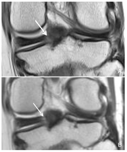
Figure 2
A 37-year-old man with focal cartilage defect and subchondral fracture in lateral tibial plateau. 2D coronal intermediate-weighted image (A) and reformatted coronal image of 3D isotropic intermediate-weighted sequence (B) of the knee demonstrate focal chondromalacia (arrow) clearly at 3 tesla magnetic resonance imaging.
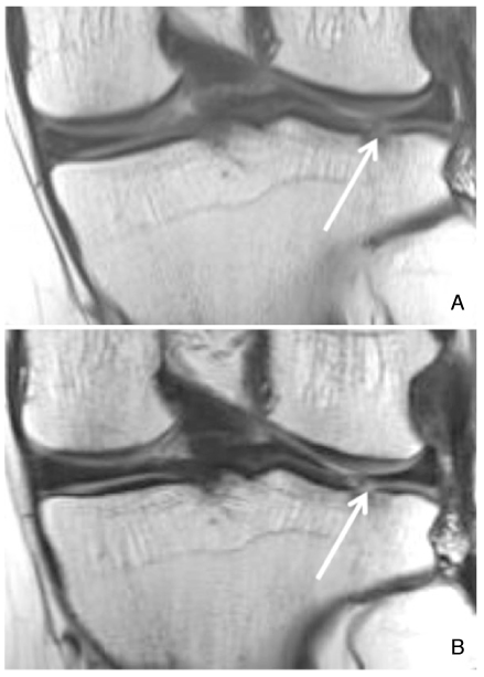
Figure 3
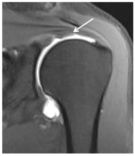
A 22-year-old woman with osteonecrosis of right femoral head and secondary osteoarthritis. Fat-suppressed, coronal intermediate-weighted image at 3 tesla magnetic resonance imaging shows well diffuse irregularity of hyaline cartilage within the hip joint due to secondary osteoarthritis.
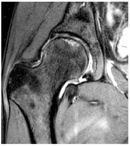
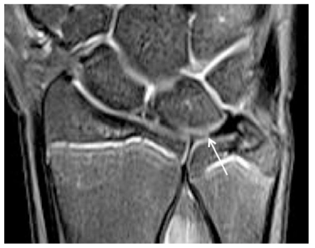
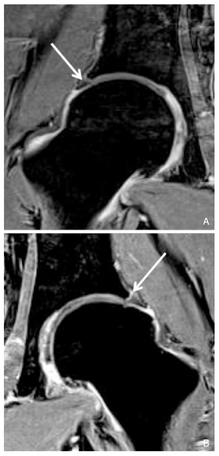
Figure 4
A 20-year-old man with healing process of subchondral insufficiency fracture at bilateral femoral heads. Coronal T1 VIBE images of both hip joints demonstrate normal acetabular labrum (arrow) within right hip joint (A) and deformed and detached labrum (arrow) of left hip joint (B).
- TOOLS
-
- Share :
-


-
METRICS

-
- 0 Crossref
- Scopus
- 1,232 View
- 4 Download
-

-
Related articles in
J Korean Med Assoc -
Health insurance policies for magnetic resonance imaging tests in Korea2021 March;64(3)
Clinical application of high field strength magnetic resonance imaging2010 December;53(12)
Introduction to high field strength magnetic resonance imaging2010 December;53(12)
High field strength magnetic resonance imaging of cardiovascular diseases2010 December;53(12)
High field strength magnetic resonance imaging of abdominal diseases2010 December;53(12)
- Editorial Office
-
37 Ichon-ro 46-gil, Yongsan-gu, Seoul
Tel: +82-2-6350-6562 Fax: +82-2-792-5208 E-mail: jkmamaster@gmail.com
Copyright © 2024 by Korean Medical Association.
