


 |
 |
| J Korean Med Assoc > Volume 52(2); 2009 > Article |
Abstract
Atherosclerosis is characterized by progressive accumulation of lipids and inflammatory cells within the artery wall. It is a diffuse systemic disease; however, some atherosclerotic plaques are more prone to rupture causing sudden thromboembolic vascular occlusions, while others are clinically silent. Therefore, to prevent such devastating vascular events as stroke or myocardial infarction, clinicians need to have smart tools to localize high-risk vulnerable plaques, which have been a huge challenge to date. Molecular imaging, which visualizes biologic processes at the cellular and molecular level, has a potential to assess plaque vulnerability and consequently identify high-risk patients prior to the development of the clinical events. In this review, we summarize important updates on the molecular imaging of atherosclerosis in the field of optical imaging, magnetic resonance imaging, positron emission tomography, and computerized tomography imaging.
References
1. Kung H. Deaths: Final Data for 2005. National Vital Statistics Reports 56.
2. Libby P. Inflammation in atherosclerosis. Nature 2002;420:868-874.
3. Rader DJ, Daugherty A. Translating molecular discoveries into new therapies for atherosclerosis. Nature 2008;451:904-913.
4. Ross R. Atherosclerosis-an inflammatory disease. N Engl J Med 1999;340:115-126.
5. Lusis AJ. Atherosclerosis. Nature 2000;407:233-241.
6. Geroulakos G, Ramaswami G, Nicolaides A, James K, Labropoulos N, Belcaro G, Holloway M. Charac-terization of symp tomatic and asymptomatic carotid plaques using highresolution real-time ultrasonography. Br J Surg 1993;80:1274-1277.
7. Grant EG, Benson CB, Moneta GL, Alexandrov AV, Baker JD, Bluth EI, Carroll BA, Eliasziw M, Gocke J, Hertzberg BS, Katanick S, Needleman L, Pellerito J, Polak JF, Rholl KS, Wooster DL, Zierler RE. Carotid artery stenosis: gray-scale and Doppler US diagnosis-Society of Radiologists in Ultrasound Consensus Conference. Radiology 2003;229:340-346.
8. Gronholdt ML. Ultrasound and lipoproteins as predictors of lipid-rich, rupture-prone plaques in the carotid artery. Arterioscler Thromb Vasc Biol 1999;19:2-13.
9. Gronholdt ML, Nordestgaard BG, Wiebe BM, Wilhjelm JE, Sillesen H. Echo-lucency of computerized ultrasound images of carotid atherosclerotic plaques are associated with increased levels of triglyceride-rich lipoproteins as well as increased plaque lipid content. Circulation 1998;97:34-40.
10. Jaffer FA, Libby P, Weissleder R. Molecular and cellular imaging of atherosclerosis: emerging applications. J Am Coll Cardiol 2006;47:1328-1338.
11. Jaffer FA, Libby P, Weissleder R. Molecular imaging of cardiovascular disease. Circulation 2007;116:1052-1061.
12. Jaffer FA, Weissleder R. Molecular imaging in the clinical arena. Jama 2005;293:855-862.
13. Weissleder R, Mahmood U. Molecular imaging. Radiology 2001;219:316-333.
14. Mayo SW, Eldrup-Jorgensen J, Lucas FL, Wennberg DE, Bredenberg CE. Carotid endarterectomy after NASCET and ACAS: a statewide study. North American Symptomatic Carotid Endarterectomy Trial. Asymptomatic Carotid Artery Stenosis Study. J Vasc Surg 1998;27:1017-1022. discussion 22-23.
15. Moore WS, Barnett HJ, Beebe HG, Bernstein EF, Brener BJ, Brott T, Caplan LR, Day A, Goldstone J, Hobson RW 2nd, et al. Guidelines for carotid endarterectomy. A multidisciplinary consensus statement from the Ad Hoc Committee, American Heart Association. Circulation 1995;91:566-579.
16. Smith SC Jr, Feldman TE, Hirshfeld JW Jr, Jacobs AK, Kern MJ, King SB 3rd, Morrison DA, O'Neill WW, Schaff HV, Whitlow PL, Williams DO, Antman EM, Adams CD, Anderson JL, Faxon DP, Fuster V, Halperin JL, Hiratzka LF, Hunt SA, Nishimura R, Ornato JP, Page RL, Riegel B. ACC/AHA/SCAI 2005 Guideline Update for Percutaneous Coronary Intervention-summary article: a report of the American College of Cardiology/American Heart Association Task Force on Practice Guidelines (ACC/AHA/SCAI Writing Committee to Update the 2001 Guidelines for Percutaneous Coronary Intervention). Circulation 2006;113:156-175.
17. Barnett HJ, Taylor DW, Eliasziw M, Fox AJ, Ferguson GG, Haynes RB, Rankin RN, Clagett GP, Hachinski VC, Sackett DL, Thorpe KE, Meldrum HE, Spence JD. Benefit of carotid endarterectomy in patients with symptomatic moderate or severe stenosis. North American Symptomatic Carotid Endarterectomy Trial Collaborators. N Engl J Med 1998;339:1415-1425.
18. Biller J, Feinberg WM, Castaldo JE, Whittemore AD, Harbaugh RE, Dempsey RJ, Caplan LR, Kresowik TF, Matchar DB, Toole JF, Easton JD, Adams HP Jr, Brass LM, Hobson RW 2nd, Brott TG, Sternau L. Guidelines for carotid endarterectomy: a statement for healthcare professionals from a Special Writing Group of the Stroke Council, American Heart Association. Circulation 1998;97:501-509.
19. Inzitari D, Eliasziw M, Gates P, Sharpe BL, Chan RK, Meldrum HE, Barnett HJ. The causes and risk of stroke in patients with asymptomatic internal-carotid-artery stenosis. North American Symptomatic Carotid Endarterectomy Trial Collaborators. N Engl J Med 2000;342:1693-1700.
20. Jaffer FA, Kim DE, Quinti L, Tung CH, Aikawa E, Pande AN, Kohler RH, Shi GP, Libby P, Weissleder R. Optical visualization of cathepsin K activity in atherosclerosis with a novel, protease-activatable fluorescence sensor. Circulation 2007;115:2292-2298.
21. Aikawa M, Libby P. The vulnerable atherosclerotic plaque: pathogenesis and therapeutic approach. Cardiovasc Pathol 2004;13:125-138.
22. Chen J, Tung CH, Mahmood U, Ntziachristos V, Gyurko R, Fishman MC, Huang PL, Weissleder R. In vivo imag-ing of proteolytic activity in atherosclerosis. Circulation 2002;105:2766-2771.
23. Deguchi JO, Aikawa M, Tung CH, Aikawa E, Kim DE, Ntziachristos V, Weissleder R, Libby P. Inflammation in atherosclerosis: visualizing matrix metalloproteinase action in macrophages in vivo. Circulation 2006;114:55-62.
24. Kim DE, Kim JY, Kim EJ, Jeong SW. Molecular Optical Imaging of Cathepsin-B Proteolytic Enzyme Activity to Reflect Atherosclerosis Pathophysiology and Anti-Atherosclerotic Therapeutic Effect. J Korean Neurol Assoc 2009;in press.
25. Moon WK, Moon WK. Molecular MR Imaging. J Korean Med Assoc 2004;47:133-138.
26. Sosnovik DE, Nahrendorf M, Weissleder R. Molecular magnetic resonance imaging in cardiovascular medicine. Circulation 2007;115:2076-2086.
27. Kooi ME, Cappendijk VC, Cleutjens KB, Kessels AG, Kitslaar PJ, Borgers M, Frederik PM, Daemen MJ, van Engelshoven JM. Accumulation of ultrasmall super-paramagnetic particles of iron oxide in human atherosclerotic plaques can be detected by in vivo magnetic resonance imaging. Circulation 2003;107:2453-2458.
28. Müller K, Skepper JN, Tang TY, Graves MJ, Patterson AJ, Corot C, Lancelot E, Thompson PW, Brown AP, Gillard JH. Atorvastatin and uptake of ultrasmall superparamagnetic iron oxide nanoparticles (Ferumoxtran-10) in human monocyte-macrophages: implications for magnetic resonance imaging. Biomaterials 2008;29:2656-2662.
29. Brooks PC, Clark RA, Cheresh DA. Requirement of vascular integrin alpha v beta 3 for angiogenesis. Science 1994;264:569-571.
30. Winter PM, Morawski AM, Caruthers SD, Fuhrhop RW, Zhang H, Williams TA, Allen JS, Lacy EK, Robertson JD, Lanza GM, Wickline SA. Molecular imaging of angiogenesis in early-stage atherosclerosis with alpha (v) beta3-integrin-targeted nanoparticles. Circulation 2003;108:2270-2274.
31. Chen JW, Pham W, Weissleder R, Bogdanov A Jr. Human myeloperoxidase: a potential target for molecular MR imaging in atherosclerosis. Magn Reson Med 2004;52:1021-1028.
32. Nahrendorf M, Jaffer FA, Kelly KA, Sosnovik DE, Aikawa E, Libby P, Weissleder R. Noninvasive vascular cell adhesion molecule-1 imaging identifies inflammatory activation of cells in atherosclerosis. Circulation 2006;114:1504-1511.
33. Chu B, Kampschulte A, Ferguson MS, Kerwin WS, Yarnykh VL, O'Brien KD, Polissar NL, Hatsukami TS, Yuan C. Hemorrhage in the atherosclerotic carotid plaque: a high-resolution MRI study. Stroke 2004;35:1079-1084.
34. Saam T, Ferguson MS, Yarnykh VL, Takaya N, Xu D, Polissar NL, Hatsukami TS, Yuan C. Quantitative evaluation of carotid plaque composition by in vivo MRI. Arterioscler Thromb Vasc Biol 2005;25:234-239.
35. Larose E, Kinlay S, Selwyn AP, Yeghiazarians Y, Yucel EK, Kacher DF, Libby P, Ganz P. Improved characterization of atherosclerotic plaques by gadolinium contrast during intravascular magnetic resonance imaging of human arteries. Atherosclerosis 2008;196:919-925.
36. Larose E, Yeghiazarians Y, Libby P, Yucel EK, Aikawa M, Kacher DF, Aikawa E, Kinlay S, Schoen FJ, Selwyn AP, Ganz P. Characterization of human atherosclerotic plaques by intravascular magnetic resonance imaging. Circulation 2005;112:2324-2331.
37. Davies JR, Rudd JH, Fryer TD, Graves MJ, Clark JC, Kirkpatrick PJ, Kooi ME, Cappendijk VC, Cleutjens KB, Kessels AG, Kitslaar PJ, Borgers M, Frederik PM, Daemen MJ, van Engelshoven JM. Identification of culprit lesions after transient ischemic attack by combined [18F]-fluorodeoxyglucose positron emission tomography and high-resolution magnetic resonance imaging. Stroke 2005;36:2642-2647.
38. Rudd JH, Warburton EA, Fryer TD, Jones HA, Clark JC, Antoun N, Johnström P, Davenport AP, Kirkpatrick PJ, Arch BN, Pickard JD, Weissberg PL. Imaging atherosclerotic plaque inflammation with [18F]-fluorodeoxyglucose positron emission tomography. Circulation 2002;105:2708-2711.
39. Tawakol A, Migrino RQ, Bashian GG, Bedri S, Vermylen D, Cury RC, Yates D, LaMuraglia GM, Furie K, Houser S, Gewirtz H, Muller JE, Brady TJ, Fischman AJ. In vivo 18F-fluorode-oxyglucose positron emission tomography imaging provides a noninvasive measure of carotid plaque inflammation in patients. J Am Coll Cardiol 2006;48:1818-1824.
40. Kircher MF, Grimm J, Swirski FK, Libby P, Gerszten RE, Allport JR, Weissleder R. Noninvasive in vivo imaging of monocyte trafficking to atherosclerotic lesions. Circulation 2008;117:388-395.
41. Hyafil F, Cornily JC, Feig JE, Gordon R, Vucic E, Amirbekian V, Fisher EA, Fuster V, Feldman LJ, Fayad ZA. Noninvasive detection of macrophages using a nanoparticulate contrast agent for computed tomography. Nat Med 2007;13:636-641.
Figure 1
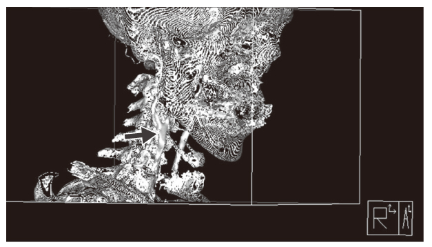
Duplex ultrasonography (A) and angiography (B) imaging to provide structural information about carotid atherosclerosis. (A) On the carotid sonography, the atherosclerotic plaque is shown to have heterogeneous echodensity(blue arrows). (B) The angiography shows significant atherosclerotic narrowing of the internal carotid artery. Current atherosclerosis practice heavily relies on these anatomy-based structural imaging modalities.
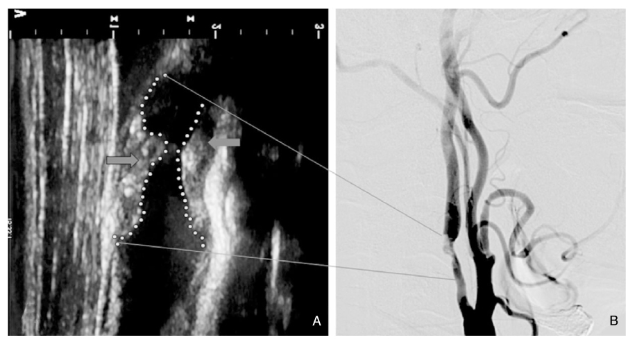
Figure 2
Cathepsin-B molecular optical imaging to reflect pro-atherosclerotic and anti-atherosclerotic effects by high cholesterol diet and atorvastatin treatment, respectively. Eight-week-old ApoE knock-out mice were on a normal chow diet, western diet, or western diet with atorvastatin for 14 weeks. The Cathepsin-B images of the representative animals' aortas show that Cathepsin-B activity signal is stronger in the mouse on a high cholesterol diet than in the mouse on a normal diet. In the animal on a western diet + atorvastatin, the Cathepsin-B activity signal is as low as in the animal on a normal diet. Please note that similarly-looking plaques on the color photograph (of the animal on a western diet) have different signal intensities on the CatB image (arrows and arrow heads). Adapted from Kim et al (24) with permission from the Korean Neurological Association.
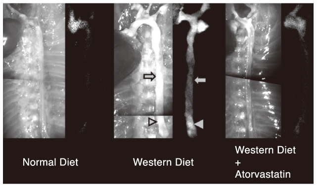
Figure 3
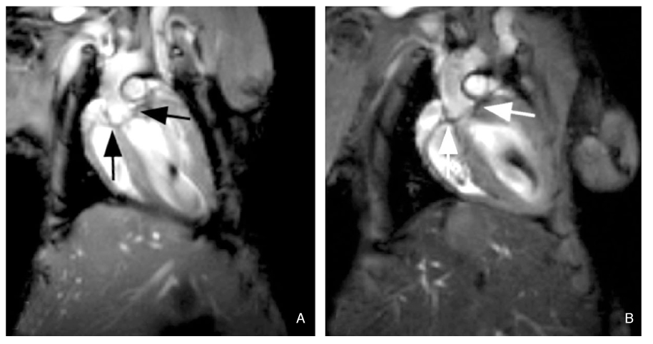
MR Images before (A) and after (B) the intravenous injection of CLIO (cross-linked iron oxide) nanoparticles into the 9-month-old ApoE k/o mouse with atherosclerotic aorta. Compared with the baseline image (A), post-contrast image (B) reveals dark signal from macrophages that phagocitized the nanoparticles and migrated to the root of the atherosclerotic aorta. (Figure courtesy of Dr. Matthias Nahrendorf, Harvard Medical School).
- TOOLS
-
- Share :
-


- Editorial Office
-
37 Ichon-ro 46-gil, Yongsan-gu, Seoul
Tel: +82-2-6350-6562 Fax: +82-2-792-5208 E-mail: jkmamaster@gmail.com
Copyright © 2024 by Korean Medical Association.
