


 |
 |
| J Korean Med Assoc > Volume 52(1); 2009 > Article |
Abstract
Spinal dysraphism is a common birth defect that causes different kinds of secondary impairments, including joint deformities, reduced mobility, and bowel or bladder dysfunction. Various dysraphic spinal abnormalities result in tethered cord syndrome, a progressive form of neurological deterioration that results from spinal cord tethering. The surgery and management of children who have spinal dysraphism require multidisciplinary care and long-term follow-up by multiple specialists in birth defects. This article reviews the clinical presentation, pathophysiology, diagnostic strategies, and therapeutic management of spinal dysraphism in infancy.
References
1. Atala A, Bauer SB, Dyro FM, Shefner J, Shillito J, Sathi S, Scott RM. Bladder functional changes resulting from lipomyelomeningocele repair. J Urol 1992;148:592-594.
2. Aufschnaiter K, Fellner F, Wurm G. Surgery in adult onset tethered cord syndrome (ATCS): review of literature on occasion of an exceptional case. Neurosurg Rev 2008.
3. Bellin MH, Sawin KJ, Roux G, Buran CF, Brei TJ. The experience of adolescent women living with spina bifida part I: self-concept and family relationships. Rehabil Nurs 2007;32:57-67.
4. Benton TJ, Cheema R, Nirgiotis JG. A newborn with a skin lesion on the back. J Fam Pract 2007;56:E1-E3.
5. Bowman RM, McLone DG, Grant JA, Tomita T, Ito JA. Spina bifida outcome: a 25-year prospective. Pediatr Neurosurg 2001;34:114-120.
6. Brand MC. Part 3: examination of the newborn with closed spinal dysraphism. Adv Neonatal Care 2007;7:30-40. quiz 2007; 41-32.
7. Brophy JD, Sutton LN, Zimmerman RA, Bury E, Schut L. Magnetic resonance imaging of lipomyelomeningocele and tethered cord. Neurosurgery 1989;25:336-340.
8. Bruner JP, Richards WO, Tulipan NB, Arney TL. Endoscopic coverage of fetal myelomeningocele in utero. Am J Obstet Gynecol 1999;180:153-158.
9. Bui CJ, Tubbs RS, Oakes WJ. Tethered cord syndrome in children: a review. Neurosurg Focus 2007;23:1-9.
10. Byrne RW, Hayes EA, George TM, McLone DG. Operative resection of 100 spinal lipomas in infants less than 1 year of age. Pediatr Neurosurg 1995;23:182-186. discussion 186-187.
11. Cabaret AS, Loget P, Loeuillet L, Odent S, Poulain P. Embryology of neural tube defects: information provided by associated malformations. Prenat Diagn 2007;27:738-742.
12. Chapman PH. Congenital intraspinal lipomas: anatomic considerations and surgical treatment. Childs Brain 1982;9:37-47.
13. Chaseling RW, Johnston IH, Besser M. Meningoceles and the tethered cord syndrome. Childs Nerv Syst 1985;1:105-108.
14. Colak A, Pollack IF, Albright AL. Recurrent tethering: a common long-term problem after lipomyelomeningocele repair. Pediatr Neurosurg 1998;29:184-190.
15. Daszkiewicz P, Barszcz S, Roszkowski M, Maryniak A. Tethered cord syndrome in children-impact of surgical treatment on functional neurological and urological outcome. Neurol Neurochir Pol 2007;41:427-435.
16. Dias MS, Walker ML. The embryogenesis of complex dysraphic malformations: a disorder of gastrulation? Pediatr Neurosurg 1992;18:229-253.
17. Finn MA, Walker ML. Spinal lipomas: clinical spectrum, embryology, and treatment. Neurosurg Focus 2007;23:1-12.
18. Ghi T, Pilu G, Falco P, Segata M, Carletti A, Cocchi G, Santini D, Bonasoni P, Tani G, Rizzo N. Prenatal diagnosis of open and closed spina bifida. Ultrasound Obstet Gynecol 2006;28:899-903.
19. Guerra LA, Pike J, Milks J, Barrowman N, Leonard M. Outcome in patients who underwent tethered cord release for occult spinal dysraphism. J Urol 2006;176:1729-1732.
20. Hahn YS. Open myelomeningocele. Neurosurg Clin N Am 1995;6:231-241.
21. Herman JM, McLone DG, Storrs BB, Dauser RC. Analysis of 153 patients with myelomeningocele or spinal lipoma reoperated upon for a tethered cord. Presentation, management and outcome. Pediatr Neurosurg 1993;19:243-249.
22. Hoffman HJ, Taecholarn C, Hendrick EB, Humphreys RP. Management of lipomyelomeningoceles. Experience at the Hospital for Sick Children, Toronto. J Neurosurg 1985;62:1-8.
23. Horton D, Barnes P, Pendleton BD, Pollay M. Spina bifida occulta: early clinical and radiographic diagnosis. J Okla State Med Assoc 1989;82:15-19.
24. Kanev PM, Lemire RJ, Loeser JD, Berger MS. Management and long-term follow-up review of children with lipomyelomeningocele, 1952-1987. J Neurosurg 1990;73:48-52.
25. Kim SY, McGahan JP, Boggan JE, McGrew W. Prenatal diagnosis of lipomyelomeningocele. J Ultrasound Med 2000;19:801-805.
26. Korner I, Schluter C, Lax H, Rubben H, Radmayr C. [Health-related quality of life in children with spina bifida]. Urologe A 2006;45:620-625.
27. La Marca F, Grant JA, Tomita T, McLone DG. Spinal lipomas in children: outcome of 270 procedures. Pediatr Neurosurg 1997;26:8-16.
28. Lemelle JL, Guillemin F, Aubert D, Guys JM, Lottmann H, Lortat-Jacob S, Moscovici J, Mouriquand P, Ruffion A, Schmitt M. A multicenter evaluation of urinary incontinence management and outcome in spina bifida. J Urol 2006;175:208-212.
29. Lemelle JL, Guillemin F, Aubert D, Guys JM, Lottmann H, Lortat-Jacob S, Mouriquand P, Ruffion A, Moscovici J, Schmitt M. Quality of life and continence in patients with spina bifida. Qual Life Res 2006;15:1481-1492.
30. Lew SM, Kothbauer KF. Tethered cord syndrome: an updated review. Pediatr Neurosurg 2007;43:236-248.
31. Lorber J. Results of treatment of myelomeningocele. An analysis of 524 unselected cases, with special reference to possible selection for treatment. Dev Med Child Neurol 1971;13:279-303.
32. Lunardi P, Missori P, Ferrante L, Fortuna A. Long-term results of surgical treatment of spinal lipomas. Report of 18 cases. Acta Neurochir (Wien) 1990;104:64-68.
33. Martinez-Lage JF, Niguez BF, Perez-Espejo MA, Almagro MJ, Maeztu C. Midline cutaneous lumbosacral lesions: not always a sign of occult spinal dysraphism. Childs Nerv Syst 2006;22:623-627.
34. McLaughlin MR, O'Connor NR, Ham P. Newborn skin: Part II. Birthmarks. Am Fam Physician 2008;77:56-60.
35. McLone DG. Technique for closure of myelomeningocele. Childs Brain 1980;6:65-73.
36. McLone DG. Results of treatment of children born with a myelomeningocele. Clin Neurosurg 1983;30:407-412.
37. McLone DG, Dias MS. Complications of myelomeningocele closure. Pediatr Neurosurg 1991;17:267-273.
38. Nelson MD Jr, Bracchi M, Naidich TP, McLone DG. The natural history of repaired myelomeningocele. Radiographics 1988;8:695-706.
39. Padmanabhan R. Etiology, pathogenesis and prevention of neural tube defects. Congenit Anom (Kyoto) 2006;46:55-67.
40. Pang D. In: Wilkins RH, Rengachary SS, editor. Tethered cord sysdrome. Neurosurgery 1996;New York: McGraw-Hill. 3465-3496.
41. Park TS. In: Albright L, Pollack I, Adelson D, editor. Myelomeningocele. Principles and Practice of Pediatric Neurosurgery 1999;New York: Thiem Medical. 291-320.
42. Perera GK, Atherton D. The value of MRI in a patient with occult spinal dysraphism. Pediatr Dermatol 2006;23:24-26.
43. Pierre-Kahn A, Lacombe J, Pichon J, Giudicelli Y, Renier D, Sainte-Rose C, Perrigot M, Hirsch JF. Intraspinal lipomas with spina bifida. Prognosis and treatment in 73 cases. J Neurosurg 1986;65:756-761.
44. Reigel DH. In: Cheek WR, Marlin AE, McLone DG, editor. Tethered spinal cord. Pediatric Neurosurgery; Surgery of the Developing Nervous System 1994;Philadelphia: WB Saunders. 77-95.
45. Schenk JP, Herweh C, Gunther P, Rohrschneider W, Zieger B, Troger J. Imaging of congenital anomalies and variations of the caudal spine and back in neonates and small infants. Eur J Radiol 2006;58:3-14.
46. Schropp C, Sorensen N, Collmann H, Krauss J. Cutaneous lesions in occult spinal dysraphism-correlation with intraspinal findings. Childs Nerv Syst 2006;22:125-131.
47. Schropp C, Speer CP, Schweitzer T, Krauss J. [Congenital skin lesions in occult spinal dysraphism-what is typical?] Z Geburtshilfe Neonatol 2006;210:222-227.
48. Schut L, Bruce DA, Sutton LN. The management of the child with a lipomyelomeningocele. Clin Neurosurg 1983;30:464-476.
49. Shurtleff DB, Lemire RJ. Epidemiology, etiologic factors, and prenatal diagnosis of open spinal dysraphism. Neurosurg Clin N Am 1995;6:183-193.
50. Steinbok P, Irvine B, Cochrane DD, Irwin BJ. Long-term outcome and complications of children born with meningomyelocele. Childs Nerv Syst 1992;8:92-96.
51. Sutton LN. Lipomyelomeningocele. Neurosurg Clin N Am 1995;6:325-338.
52. Tamaki N, Shirataki K, Kojima N, Shouse Y, Matsumoto S. Tethered cord syndrome of delayed onset following repair of myelomeningocele. J Neurosurg 1988;69:393-398.
53. Tubbs RS, Oakes WJ. A simple method to deter retethering in patients with spinal dysraphism. Childs Nerv Syst 2006;22:715-716.
54. Tulipan N, Hernanz-Schulman M, Bruner JP. Reduced hindbrain herniation after intrauterine myelomeningocele repair: A report of four cases. Pediatr Neurosurg 1998;29:274-278.
55. Williams H. A unifying hypothesis for hydrocephalus, Chiari malformation, syringomyelia, anencephaly and spina bifida. Cerebrospinal Fluid Res 2008;5:7.
56. Wills KE, Holmbeck GN, Dillon K, McLone DG. Intelligence and achievement in children with myelomeningocele. J Pediatr Psychol 1990;15:161-176.
57. Zide BM. How to reduce the morbidity of wound closure following extensive and complicated laminectomy and tethered cord surgery. Pediatr Neurosurg 1992;18:157-166.
Figure 1
Diagram of spina bifida and MR axial view. (A, E): normal, (B): spina bifida occulta, (C, F): spina bifida cytica-meningocele, (D, G): spina bifida cytica-myelomeningocele.
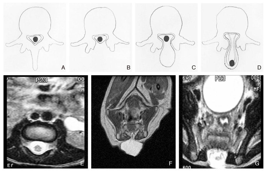
Figure 2
Spina bifida, open.
(A) Myeloschisis, (B) Spina bifida cystica-meningocle, (C) Spina bifida cystica-myelomeningocle.

Figure 3
Various dermatologic feature in closed spina bifida.
(A) Lipoma, (B) Skin depression, (C) Capillary telangiectasis.
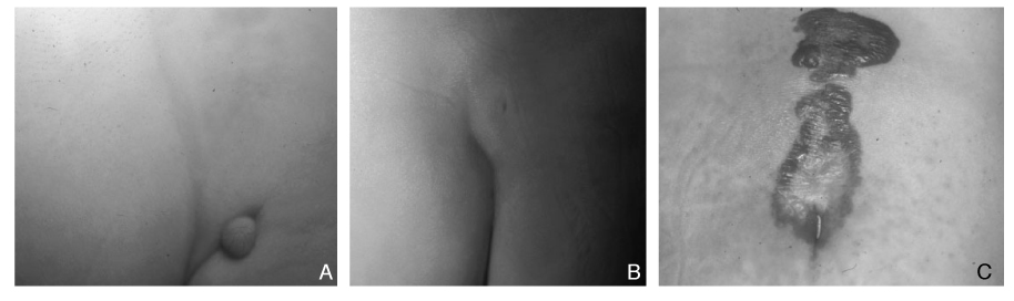
Figure 4
Location of conus medullaris.
(A) Postovulation 8 wks, (B) Postovulation 24 wks, (C) Birth, (D) Normal adult.

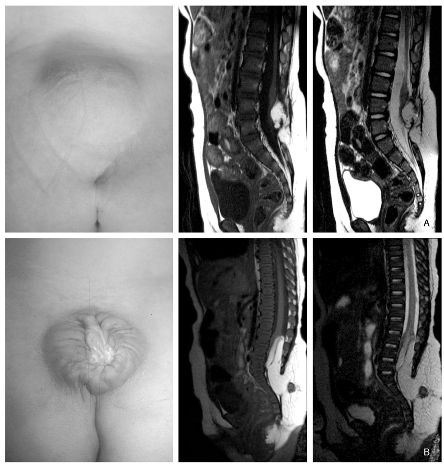
Figure 7
Fetal sonography (left) and fetal MRI (right) showing mylelomeningocele at post gestation 22wks.
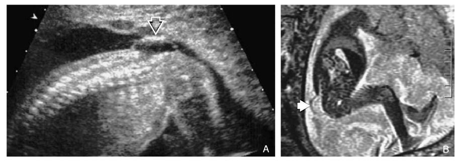
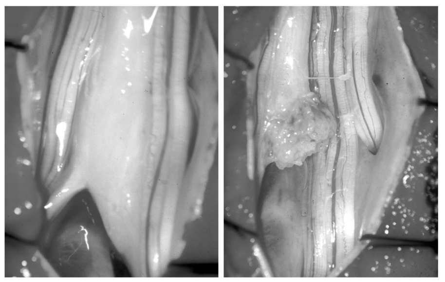
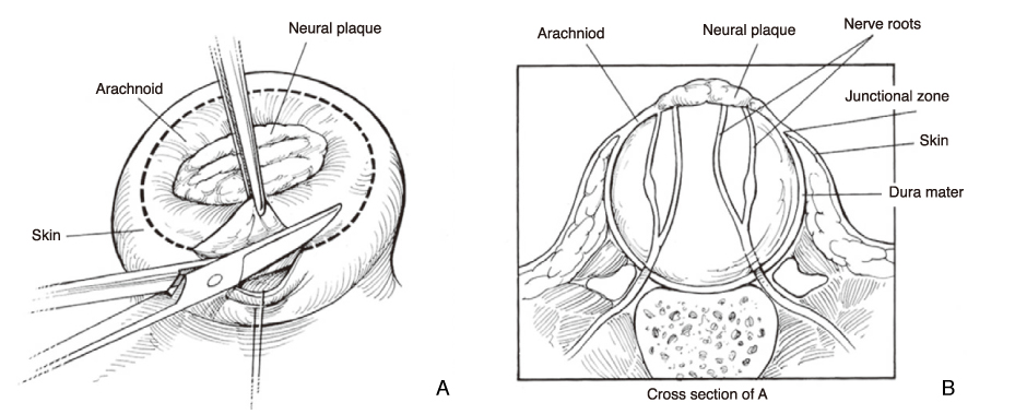
Figure 9
(A) Initial incision follows the circumferential white line formed by the junction of the arachnoid and skin, (B) Cross section of A.
- TOOLS
-
- Share :
-


-
METRICS

-
- 0 Crossref
- Scopus
- 1,324 View
- 8 Download
-

-
Related articles in
J Korean Med Assoc -
Diagnosis and Treatment of Irritable Bowel Syndrome2003 July;46(7)
Diagnosis and management of acute coronary syndrome2017 July;60(7)
- Editorial Office
-
37 Ichon-ro 46-gil, Yongsan-gu, Seoul
Tel: +82-2-6350-6562 Fax: +82-2-792-5208 E-mail: jkmamaster@gmail.com
Copyright © 2024 by Korean Medical Association.
