


 |
 |
| J Korean Med Assoc > Volume 52(1); 2009 > Article |
Abstract
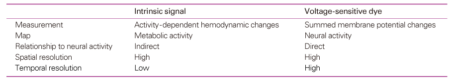
Functional mapping techniques including functional magnetic resonance imaging (fMRI), positron emission tomography (PET), and magnetoencephalography (MEG) can be used to study the function of the nervous system. Optical imaging is an emerging technique for functional imaging of the nervous tissue. Functional optical imaging can be classified into two major streams; intrinsic signal optical imaging (ISO) and voltage-sensitive dye optical imaging (VDO). ISO is related to hemodynamic changes such as hemoglobin concentration and oxygenation changes, cytochrome oxidation change, and light scattering. On the contrary, VOD measures changes in membrane potentials of neural cells. Therefore, ISO reflects metabolic activity of neurons, while VOD directly reflects neural activity. Recent advances in optical imaging opened the possibility of its application to clinical situations as well as basic researches. Further, development of optical imaging may greatly contribute to the understanding of the function of the nervous system.
References
1. Baker BJ, Kosmidis EK, Vucinic D, Falk CX, Cohen LB, Djurisic M, Zecevic D. Imaging brain activity with voltage-and calcium-sensitive dyes. Cell Mol Neurobiol 2005;25:245-282.
2. Pouratian N, Cannestra AF, Martin NA, Toga AW. Intraoperative optical intrinsic signal imaging: a clinical tool for functional brain mapping. Neurosurg Focus 2002;13:1-9.
3. Prakash N, Uhlemann F, Sheth SA, Bookheimer S, Martin N, Toga AW. Current trends in intraoperative optical imaging for functional brain mapping and delineation of lesions of language cortex. Neuroimage 2008;(in press).
4. Aitken PG, Fayuk D, Somjen GG, Turner DA. Use of intrinsic optical signals to monitor physiological changes in brain tissue slices. Methods 1999;18:91-103.
5. Roe AW. Long-term optical imaging of intrinsic signals in anesthetized and awake monkeys. Appl Opt 2007;46:1872-1880.
6. Grinvald A, Hildesheim R. VSDI: a new era in functional imaging of cortical dynamics. Nat Rev Neurosci 2004;5:874-885.
7. Mrsic-Flogel T, Hübener M, Bonhoeffer T. Brain mapping: new wave optical dispatch imaging. Curr Biol 2003;13:R778-R780.
8. Hill DK, Keynes RD. Opacity changes in stimulated nerve. J Physiol 1949;108:278-281.
9. Narayan SM, Santori EM, Blood AJ, Burton JS, Toga AW. Imaging optical reflectance in rodent barrel and forelimb sensory cortex. Neuroimage 1994;1:181-190.
10. Bonhoeffer T, Grinvald A. Iso-orientation domains in cat visual cortex are arranged in pinwheel-like patterns. Nature 1991;353:429-431.
11. Frostig RD, Lieke EE, Ts'o DY, Grinvald A. Cortical functional architecture and local coupling between neuronal activity and the microcirculation revealed by in vivo high-resolution optical imaging of intrinsic signals. Proc Natl Acad Sci USA 1990;87:6082-6086.
12. Grinvald A, Frostig RD, Siegel RM, Bartfeld E. High-resolution optical imaging of functional brain architecture in the awake monkey. Proc Natl Acad Sci USA 1991;88:11559-11563.
13. Haglund MM, Ojemann GA, Blasdel GG. Optical imaging of bipolar cortical stimulation. J Neurosurg 1993;78:785-793.
14. Sasaki S, Yazawa I, Miyakawa N, Mochida H, Shinomiya K, Kamino K, Momose-Sato Y, Sato K. Optical imaging of intrinsic signals induced by peripheral nerve stimulation in the in vivo rat spinal cord. Neuroimage 2002;17:1240-1255.
15. Haglund MM, Ojemann GA, Hochman DW. Optical imaging of epileptiform and functional activity in human cerebral cortex. Nature 1992;358:668-671.
16. Cannestra AF, Pouratian N, Shomer MH, Toga AW. Refractory periods observed by intrinsic signal and fluorescent dye imaging. J Neurophysiol 1998;80:1522-1532.
17. Cannestra AF, Bookheimer SY, Pouratian N, O'Farrell A, Sicotte N, Martin NA, Becker D, Rubino G, Toga AW. Temporal and topographical characterization of language cortices using intraoperative optical intrinsinc signals. Neuroimage 2000;12:41-54.
18. Pouratian N, Bookheimer SY, O'Farrell AM, Sicotte NL, Cannestra AF, Becker D, Toga AW. Optical imaging of bilingual cortical representations. Case report. J Neurosurg 2000;93:676-681.
19. Cohen LB, Salzberg BM, Davila HV, Ross WN, Landowne D, Waggoner AS, Wang CH. Changes in axon fluorescence during activity: molecular probes of membrane potential. J Membr Biol 1974;19:1-36.
20. Waggoner AS, Grinvald A. Mechanisms of rapid optical changes of potential sensitive dyes. Ann N Y Acad Sci 1977;303:217-242.
21. Waggoner AS. Dye indicators of membrane potential. Annu Rev Biophys Bioeng 1979;8:47-63.
22. Tasaki I, Watanabe A, Sandlin R, Carnay L. Changes in fluorescence, turbidity and birefringence associated with nerve excitation. Proc Natl Acad Sci USA 1968;61:883-888.
23. Salzberg BM, Davila HV, Cohen LB. Optical recording of impulses in individual neurons of an invertebrate central nervous system. Nature 1973;246:508-509.
24. Salzberg BM, Grinvald A, Cohen LB, Davila HV, Ross WN. Optical recording of neuronal activity in an invertebrate central nervous system: simultaneous recording from several neurons. J Neurophysiol 1977;40:1281-1291.
25. Tasaki I, Warashina A. Dye-membrane interaction and its changes during nerve excitation. Photochem Photobiol 1976;24:191-207.
26. Grinvald A, Cohen LB, Lesher S, Boyle MB. Simultaneous optical monitoring of activity of many neurons in invertebrate ganglia, using a 124 element photodiode array. J Neurophysiol 1981a;45:829-840.
27. Grinvald A, Ross WN, Farber I. Simultaneous optical measurements of electrical activity from multiple sites on processes of cultured neurons. Proc Natl Acad Sci USA 1981b;78:3245-3249.
28. Grinvald A, Manker A, Segal M. Visualization of the spread of electrical activity in rat hippocampal slices by voltage sensitive optical probes. J Physiol 1982;333:269-291.
29. Orbach HS, Cohen LB. Simultaneous optical monitoring of activity from many areas of the salamander olfactory bulb. A new method for studying functional organization in the vertebrate CNS. J Neurosci 1983;3:2251-2262.
30. Petersen CCH, Grinvald A, Sakmann B. Spatiotemporal dynamics of sensory responses in layer 2/3 of rat barrel cortex measured in vivo by voltage-sensitive dye imaging combined with whole-cell voltage recordings and neuron reconstructions. J Neurosci 2003;23:1298-1309.
31. Petersen CH, Grinvald A, Sakmann B. Spatio-temporal dynamics of sensory responses in layer 2/3 of rat barrel cortex measured in vivo by voltage-sensitive dye imaging combined with whole-cell voltage recordings and anatomical reconstructions. J Neurosci 2003;23:1298-1309.
32. Kaltenbach JA, Zhang JS. In vivo optical imaging of tone-evoked activity in the dorsal cochlear nucleus with a voltage sensitive dye. J Neurosci Res 2004;78:908-917.
33. Hosokawa Y, Sugimoto S, Kubota M, Taniguchi I, Horikawa J. Optical imaging of binaural interaction in multiple fields of the guinea pig auditory cortex. Neuroreport 2004;15:1093-1097.
34. Onimaru H, Homma I. A novel functional neuron group for respiratory thythm generation in the ventral medulla. J Neurosci 2003;23:1478-1486.
35. Onimaru H, Homma I. Optical imaging of respiratory neuron activity from the dorsal view of the lower brainstem. Clin Exp Pharmacol Physiol 2005;32:297-301.
36. Sato K, Nariai T, Tanaka Y, Maehara T, Miyakawa N, Sasaki S, Momose-Sato Y, Ohno K. Functional representation of the finger and face in the human somatosensory cortex: intraoperative intrinsic optical imaging. Neuroimage 2005;25:1292-1301.
Figure 1
Spatio-temporal characteristics of methodological tools in neuroscience. The property of each technique are depicted by the colored rectangles. Abscissa and ordinate indicate temporal and spatial resolution, respectively. Optical imaging covers almost the entire area (including voltage-sensitive dye imaging, intrinsic signal imaging, ion imaging, confocal imaging, multi-photon imaging, etc.). EEG, electroencephalography; fMRI, functional MRI; MEG, magnetoencephalography; PET, positron emission tomography; 2DG, 2-deoxyglucose autoradiography. (with kind permission from Grinvald and Hildesheim, 2004)
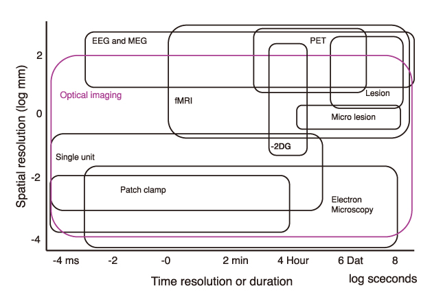
Figure 2
Voltage-sensitive dye imaging from the somatosensory cortex of the rat following electrical stimulation of the sciatic nerve.
(A) Optical imaging of cortical activation and reconstruction of a waveform.
(B) Sequential images of cortical activation following stimulation of the sciatic nerve (from upper left to lower right). Scale bar: 200µm.

- TOOLS
-
- Share :
-


-
METRICS

-
- 0 Crossref
- Scopus
- 1,122 View
- 6 Download
-

-
Related articles in
J Korean Med Assoc
- Editorial Office
-
37 Ichon-ro 46-gil, Yongsan-gu, Seoul
Tel: +82-2-6350-6562 Fax: +82-2-792-5208 E-mail: jkmamaster@gmail.com
Copyright © 2024 by Korean Medical Association.
