


 |
 |
| J Korean Med Assoc > Volume 51(12); 2008 > Article |
Abstract
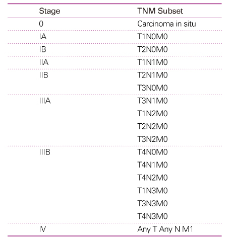
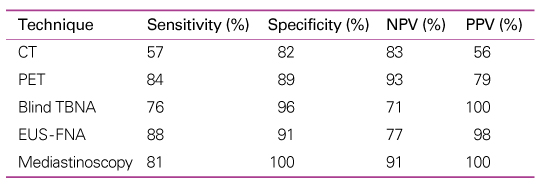
Accurate staging of lung cancers is important to determine the treatment options and the prognosis of patients with a lung cancer. TNM system revised in 1997 by American Joint Committee on Cancer and the Union Internationale Contre le Cancer is widely used in staging of the lung cancer. The TNM system is an expression of the anatomic extent of diseases and is based on the assessment of three components; extent of the primary tumor (T), regional lymph node metastasis (N), and distant metastasis (M). Non-invasive staging of lung cancers is based primarily on chest computed tomography (CT), and if available, on positron emission tomography (PET). Chest CT scanning is useful in providing anatomic details, but the accuracy of the chest CT scanning in differentiating benign from malignant lymph nodes in the mediastinum is poor. PET scanning has a much better sensitivity and specificity than chest CT scanning for mediastinal lymph node staging, and distant metastatic diseases can be detected by PET scanning. With either test, abnormal findings must be confirmed by a tissue biopsy to ensure accurate staging. Invasive techniques for biopsy of mediastinal lymph nodes or pathologic tissue include transbronchial needle aspiration, transesophageal fine needle aspiration, and surgery.
References
1. Mountain C. Revisions in the International System for Staging Lung Cancer. Chest 1997;111:1710-1717.
2. Silvestri GA, Tanoue LT, Margolis ML, Barker J, Detterbeck F. The noninvasive staging of non-small cell lung cancer: the guidelines. Chest 2003;123:147S-156S.
3. Rami-Porta R, Ball D, Crowley J, Giroux DJ, Jett J, Travis WD, Tsuboi M, Vallières E, Goldstraw P. International Staging Committee. Cancer Research and Biostatistics, Observers to the Committee, Participating Institutions. The IASLC Lung Cancer Staging Project: proposal for the revision of the T descriptors in the forth coming (seventh) edition of the TNM classification for lung cancer. J Thorac Oncol 2007;2:593-602.
4. Rusch VW, Crowley J, Giroux DJ, Goldstraw P, Im JG, Tsuboi M, Tsuchiya R, Vansteenkiste J. International Staging Committee. Cancer Research and Biostatistics, Observers to the Committee, Participating Institutions. The IASLC Lung Cancer Staging Project: proposal for the revision of the N descriptors in the forth coming (seventh) edition of the TNM classification for lung cancer. J Thorac Oncol 2007;2:603-612.
5. Lardinois D, Weder W, Hany TF, Kamel EM, Korom S, Seifert B, von Schulthess GK, Steinert HC. Staging of non-small-cell lung cancer with integrated position-emission tomography and computed tomography. N Engl J Med 2003;348:2500-2507.
6. Silvestri GA, Gould MK, Margolis ML, Lardinois D, Weder W, Hany TF, Kamel EM, Korom S, Seifert B, von Schulthess GK, Steinert HC. Noninvasive staging of non-small cell lung cancer ACCP Evidence-based clinical practice guidelines, 2nd ed. Chest 2007;132:178S-201S.
7. MacDonald SL, Hansell DM. Staging of non-small cell lung cancer: imaging of intrathoracic disease. Eur J Radiol 2003;45:18-30.
8. Weder W. Lung cancer: new opportunities-changing algorithm in staging. Ann Oncol 2008;19:28S-30S.
Table 2
International staging system TNM classification (1)

*The uncommon superficial tumor of any size with its invasive component limited to the bronchial wall, which may extend proximal to the main bronchus, is also classified T1.
†Most pleural effusions associated with lung cancer are due to tumor. However, there are a few patients in whom multiple cytopathologic examinations of pleural fluid show no tumor. In these cases, the fluid is non-bloody and is not an exudate. When these elements and clinical judgment dictate that the effusion is not related to the tumor, the effusion should be excluded as a staging element and the patient's disease should be staged T1, T2, or T3. Pericardial effusion is classified according to the same rules.
‡Separate metastatic tumor nodule (s) in the ipsilateral nonprimary-tumor lobe(s) of the lung also are classified M1.
- TOOLS
-
- Share :
-


- Editorial Office
-
37 Ichon-ro 46-gil, Yongsan-gu, Seoul
Tel: +82-2-6350-6562 Fax: +82-2-792-5208 E-mail: jkmamaster@gmail.com
Copyright © 2024 by Korean Medical Association.
