


 |
 |
| J Korean Med Assoc > Volume 50(12); 2007 > Article |
Abstract
Moyamoya disease is characterized by bilateral stenosis or occlusion of distal internal carotid artery (ICA) bifurcation including its proximal branches and abnormal vascular network (moyamoya vessel, MMV) in the vicinity of the arterial occlusions. It is the most common pediatric cerebrovascular disease in Eastern Asia, particularly in Korea and Japan. The etiology is still unknown, but much about the pathology from autopsies, factors involved in its pathogenesis, and its genetics have been studied and reported. It may cause ischemic attacks or cerebral infarctions in children and cerebral hemorrhage in adults. Because of its aggressive clinical course in very young children, the need for early detection and treatment has been recognized. Magnetic resonance imaging (MRI)/MR angiography (MRA), cerebral hemodynamic studies, and cerebral angiography are used for the diagnosis. The treatment basically focuses on prevention of further ischemia and infarction through revascularization. Technically, direct and indirect bypass methods are used. The treatment strategy needs to be individualized in each patient. Outcomes of revascularization procedures are excellent in preventing transient ischemic attacks (TIAs) in most patients.
References
1. Suzuki J, Takaku A. Cerebrovascular "moyamoya" disease. Disease showing abnormal net-like vessels in base of brain. Arch Neurol 1969;20:288-299.
2. Natori Y, Ikezaki K, Matsushima T, Fukui M. 'Angiographic moyamoya' its definition, classification, and therapy. Clin Neurol Neurosurg 1997;99:S2. S168-S172.
3. Ikezaki K, Han DH, Kawano T, Kinukawa N, Fukui M. A clinical comparison of definite moyamoya disease between South Korea and Japan. Stroke 1997;28:2513-2517.
4. Suzuki J, Kodama N. Moyamoya disease-a review. Stroke 1983;14:104-109.
5. Nakashima H, Meguro T, Kawada S, Hirotsune N, Ohmoto T. Long-term results of surgically treated moyamoya disease. Clin Neurol Neurosurg 1997;99:S2. S156-S161.
6. Ikezaki K. In: Ikezaki K, Loftus C, editor. Clinical manifestations: epidemiology, symptoms and signs, laboratory findings. Moyamoya Disease 2001;Rolling Meadows: American Association of Neurological Surgeons. 43-54.
7. Wakai K, Tamakoshi A, Ikezaki K, Fukui M, Kawamura T, Aoki R, Kojima M, Lin Y, Ohno Y. Epidemiological features of moyamoya disease in Japan: findings from a nationwide survey. Clin Neurol Neurosurg 1997;99:S2. S1-S5.
8. Kim SK, Wang KC, Kim DG, Paek SH, Chung HT, Han MH, Ahn Y, Cho BK. Clinical feature and outcome of pediatric cerebrovascular disease: a neurosurgical series. Childs Nerv Syst 2000;16:421-428.
9. Fukui M, Kono S, Sueishi K, Ikezaki K. Moyamoya disease. Neuropathology 2000;20:S2. S61-S64.
10. Malek AM, Connors S, Robertson RL, Folkman J, Scott RM. Elevation of cerebrospinal fluid levels of basic fibroblast growth factor in moyamoya and central nervous system disorders. Pediatr Neurosurg 1997;27:182-189.
11. Hojo M, Hoshimaru M, Miyamoto S, Taki W, Nagata I, Asahi M, Matsuura N, Ishizaki R, Kikuchi H, Hashimoto N. Role of transforming growth factor-beta1 in the pathogenesis of moyamoya disease. J Neurosurg 1998;89:623-629.
12. Kim SK, Yoo JI, Cho BK, Hong SJ, Kim YK, Moon JA, Kim JH, Chung YN, Wang KC. Elevation of CRABP-I in the cerebrospinal fluid of patients with Moyamoya disease. Stroke 2003;34:2835-2841.
13. Yamada H, Deguchi K, Tanigawara T, Takenaka K, Nishimura Y, Shinoda J, Hattori T, Andoh T, Sakai N. The relationship between moyamoya disease and bacterial infection. Clin Neurol Neurosurg 1997;99:S2. S221-S224.
14. Ikezaki K, Kono S, Fukui M. In: Ikezaki K, Loftus C, editor. Etiology of moyamoya disease: pathology, pathophysiology, and genetics. Moyamoya Disease 2001;Rolling Meadows: American Association of Neurological Surgeons. 55-64.
15. Kang HS, Kim SK, Cho BK, Kim YY, Hwang YS, Wang KC. Single nucleotide polymorphisms of tissue inhibitor of metalloproteinase genes in familial moyamoya disease. Neurosurgery 2006;58:1074-1080.
16. Kim SK, Seol HJ, Cho BK, Hwang YS, Lee DS, Wang KC. Moyamoya disease among young patients: its aggressive clinical course and the role of active surgical treatment. Neurosurgery 2004;54:840-844. discussion 4-6.
17. Ogawa A, Nakamura N, Yoshimoto T, Suzuki J. Cerebral blood flow in moyamoya disease. Part 2: Autoregulation and CO2 response. Acta Neurochir (Wien) 1990;105:107-111.
18. Seol HJ, Wang KC, Kim SK, Hwang YS, Kim KJ, Cho BK. Headache in pediatric moyamoya disease: review of 204 consecutive cases. J Neurosurg 2005;103:439-442.
19. Ikezaki K, Fukui M, Inamura T, Kinukawa N, Wakai K, Ono Y. The current status of the treatment for hemorrhagic type moyamoya disease based on a 1995 nationwide survey in Japan. Clin Neurol Neurosurg 1997;99:S2. S183-S186.
20. Saeki N, Nakazaki S, Kubota M, Yamaura A, Hoshi S, Sunada S, Sunami K. Hemorrhagic type moyamoya disease. Clin Neurol Neurosurg 1997;99:S2. S196-S201.
21. Iwama T, Morimoto M, Hashimoto N, Goto Y, Todaka T, Sawada M. Mechanism of intracranial rebleeding in moyamoya disease. Clin Neurol Neurosurg 1997;99:S2. S187-S190.
22. Houkin K, Yoshimoto T, Kuroda S, Ishikawa T, Takahashi A, Abe H. Angiographic analysis of moyamoya disease-how does moyamoya disease progress? Neurol Med Chir (Tokyo) 1996;36:783-787. discussion 8.
23. Kuroda S, Kamiyama H, Isobe M, Houkin K, Abe H, Mitsumori K. Cerebral hemodynamics and "re-build-up" phenomenon on electroencephalogram in children with moyamoya disease. Childs Nerv Syst 1995;11:214-219.
24. Ikezaki K, Matsushima T, Kuwabara Y, Suzuki SO, Nomura T, Fukui M. Cerebral circulation and oxygen metabolism in childhood moyamoya disease: a perioperative positron emission tomography study. J Neurosurg 1994;81:843-850.
25. Kim SK, Wang KC, Oh CW, Kim IO, Lee DS, Song IC, Cho BK. Evaluation of cerebral hemodynamics with perfusion MRI in childhood moyamoya disease. Pediatr Neurosurg 2003;38:68-75.
26. Kuwabara Y, Ichiya Y, Sasaki M, Yoshida T, Masuda K, Matsushima T, Fukui M. Response to hypercapnia in moyamoya disease. Cerebrovascular response to hypercapnia in pediatric and adult patients with moyamoya disease. Stroke 1997;28:701-707.
27. Seol HJ, Wang KC, Kim SK, Lee CS, Lee DS, Kim IO, Cho BK. Unilateral (probable) moyamoya disease: long-term follow-up of seven cases. Childs Nerv Syst 2006;22:145-150.
28. Okada Y, Shima T, Matsumura S, Nishida M, Yamada T, Okita S. [Pathophysiological studies in moyamoya disease by rCBF and cortical artery pressure measurements in comparison to those in ICA or MCA occlusion]. No To Shinkei 1988;40:899-903.
29. Matsushima Y, Inaba Y. Moyamoya disease in children and its surgical treatment. Introduction of a new surgical procedure and its follow-up angiograms. Childs Brain 1984;11:155-170.
30. Kim SK, Wang KC, Kim IO, Lee DS, Cho BK. Combined encephaloduroarteriosynangiosis and bifrontal encephalogaleo (periosteal)synangiosis in pediatric moyamoya disease. Neurosurgery 2002;50:88-96.
31. Kim CY, Wang KC, Kim SK, Chung YN, Kim HS, Cho BK. Encephaloduroarteriosynangiosis with bifrontal encephalogaleo (periosteal) synangiosis in the pediatric moyamoya disease: the surgical technique and its outcomes. Childs Nerv Syst 2003;19:316-324.
32. Chung YN, Wang KC, Cho BK. In: Kim DG, editor. Moyamoya disease, Encephaloduroarteriosynangiosis. Practical Points in Neurosurgery 2007;Seoul: Ilchokak. 385-389.
33. Kim SK, Wang KC, Cho BK. In: Kim DG, editor. Moyamoya disease, Bifrontal Encephalogaleo (periosteal) synangiosis. Practical Points in Neurosurgery 2007;Seoul: Ilchokak. 390-394.
34. Choi IJ, Hong SH, Cho BK, Wang KC, Kim SK. Encephalo-duro-arterio-synangiosis (EDAS) using occipital artery in children with moyamoya disease. J Korean Neurosurg Soc 2005;38:413-418.
35. Sainte-Rose C, Oliveira R, Puget S, Beni-Adani L, Boddaert N, Thorne J, Wray A, Zerah M, Bourgeois M. Multiple bur hole surgery for the treatment of moyamoya disease in children. J Neurosurg 2006;105:437-443.
36. Karasawa J, Touho H, Ohnishi H, Miyamoto S, Kikuchi H. Long-term follow-up study after extracranial-intracranial bypass surgery for anterior circulation ischemia in childhood moyamoya disease. J Neurosurg 1992;77:84-89.
Figure 1
An axial T2-weighted MR image shows diminished flow voids in the internal carotid and middle cerebral arteries (A) and huge cortical infarction in the right hemisphere and left frontal lobe (B). An axial T1-weighted MR image demonstrates punctuate and curvilinear flow voids of the hypertrophied moyamoya collateral in the basal ganglia (C). An axial T1-weighted MR image with gadolinium enhancement reveals prominent leptomeningeal enhancement in the right hemisphere (D).
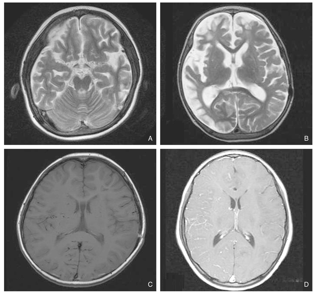
Figure 2
Rest (A) and acetazolamide (B) SPECT images show decreased perfusion with disturbed vasoreactivity to the acetazolamide injection in the right hemisphere.
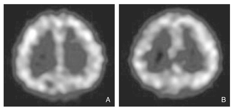
Figure 3
A perfusion MRI demonstrates an increased rCBV (A) and delayed TTP (B) in the right hemisphere.
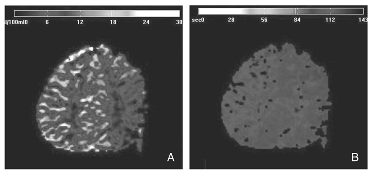
Figure 4
A right carotid angiography (A: AP view, B: lateral view) shows stenotic change in the terminal portion of the internal carotid artery, total occlusion in the middle cerebral artery and stenotic change in the proximal anterior cerebral artery. A left carotid angiography (C: AP view, D: lateral view) reveals stenotic change in the terminal portion of the internal carotid artery and the middle cerebral artery, and total occlusion in the anterior cerebral artery.
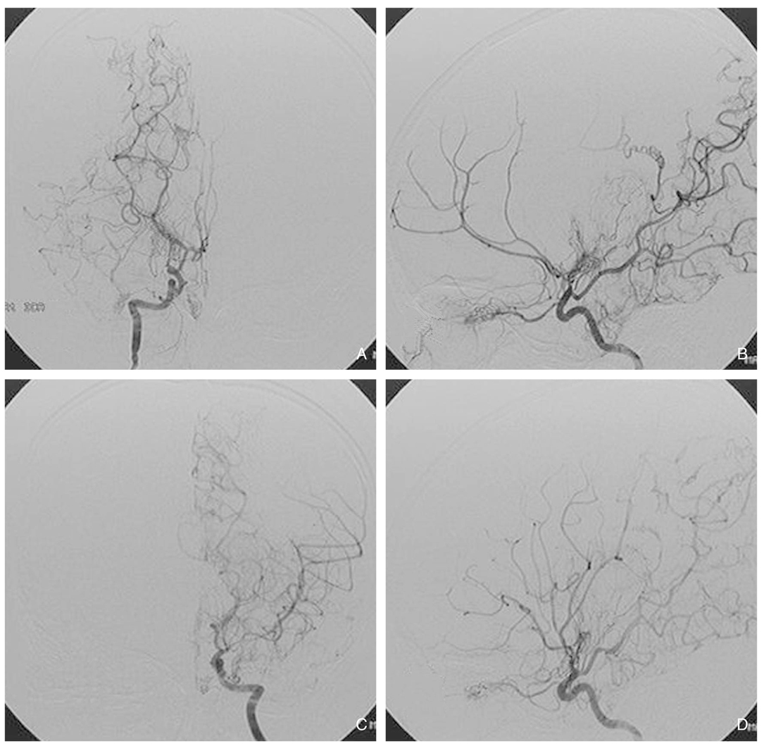
Figure 5
Operative illustrations of EDAS operation.
A) The skin incision was made along the course of the right STA.
B) The STA-galeal flap laid on the exposed cortex was sutured to the incised edge of the dura mater after dissection of the arachnoid membrane (Modified from (33) with the permission from Ilchokak).
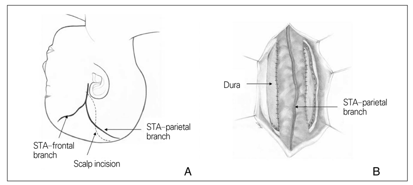
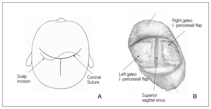
Figure 6
Operative illustrations of bifrontal EG(P)S.
A) S-shaped scalp incision was made 2 cm anterior to the coronal suture.
B) The prepared galeo (-periosteal) flap was inserted into the cerebral cortex and sutured to the dura (Modified from (35) with the permission from Ilchokak).
- TOOLS
-
- Share :
-


-
METRICS

-
- 1 Crossref
- Scopus
- 1,222 View
- 21 Download
-

-
Related articles in
J Korean Med Assoc -
Pathogenesis of HIV Disease1997 December;40(12)
Update of Radiological Diagnosis for Abdominal Disease1998 February;41(2)
- Editorial Office
-
37 Ichon-ro 46-gil, Yongsan-gu, Seoul
Tel: +82-2-6350-6562 Fax: +82-2-792-5208 E-mail: jkmamaster@gmail.com
Copyright © 2024 by Korean Medical Association.
