


 |
 |
| J Korean Med Assoc > Volume 50(2); 2007 > Article |
Abstract
The imaging technique that can provide detailed information on the left ventricular function, myocardial perfusion and viability at the same time will not only be helpful for the prognostic assessment of patients with ischemic heart disease but also be valuable in choosing appropriate therapeutic strategies. In recent years, multidetector CT (MDCT) has been proposed as a useful non-invasive imaging method of evaluating both coronary artery stenoses and cardiac morphology at the same time. MDCT has proved to be in excellent agreement with echocardiography and magnetic resonance imaging in the assessment of the left ventricular function. In addition, MDCT can provide information on myocardial viability, which can be assessed from the left ventricular wall thickness, myocardial perfusion, and a delayed contrast enhancement pattern. Despite several shortcomings to be the first-line modality for the assessment of ischemic heart disease, MDCT can provide valuable additional dynamic information in patients undergoing MDCT coronary angiography.
References
1. Park JM, Choe YH, Chang S, Sung YM, Kang SS, Kim MJ, Han BK, Choi SH. Usefulness of multidetector-row CT in the evaluation of reperfused myocardial infarction in a rabbit model. Korean J Radiol 2004;5:19-24.
2. Shah PK, Maddahi J, Staniloff HM, Ellrodt AG, Pichler M, Swan HJ, Berman DS. Variable spectrum and prognostic implications of left and right ventricular ejection fraction in patients with and without clinical heart failure after acute myocardial infarction. Am J Cardiol 1986;58:387-393.
3. White HD, Norris RM, Brown MA, Brandt PWT, Whitlock RML, Wild CJ. Left ventricular end-systolic volume as the major determination of survival after recovery from myocardial infarction. Circulation 1987;76:44-51.
4. Gerber TC, Behrenbeck T, Allison T, Mullan BP, Rumberger JA, Gibbons RJ. Comparison of measurement of left ventricular ejection fraction by Tc-99m sestamibi first-pass angiography with electron beam computed tomography in patients with anterior wall acute myocardial infarction. Am J Cardiol 1999;83:1022-1026.
5. Seter RM, Fischer SE, Lorenz CH. Quantification of left ventricular function with magnetic resonance images acquired in real time. J Magn Reson Imaging 2000;12:430-438.
6. Spuentrup E, Schroeder J, Mahnken AH, Schaeffter T, Botnar RM, Kuhl HP, Hanrath P, Gunther RW, Buecker A. Quantitative assessment of left ventricular function with interactive real-time spiral and radial MR imaging. Radiology 2003;227:870-876.
7. Nieman K, Cademartiri F, Lemos PA, Raaijmakers R, Pattynama PM, de Feyter PJ. Reliable noninvasive coronary angiography with fast submillimeter multislice spiral computed tomography. Circulation 2002;106:2051-2054.
8. Ropers D, Baum U, Pohle K, Anders K, Ulzheimer S, Ohnesorge B, Schlundt C, Bautz W, Daniel WG, Achenbach S. Detection of coronary artery stenoses with thin-slice multidetector row spiral computed tomography and multi-planar reconstruction. Circulation 2003;107:664-666.
9. Nikolaou K, Knez A, Rist C, Wintersperger BJ, Leber A, Johnson T, Reiser MF, Becker CR. Accuracy of 64-MDCT in the diagnosis of ischemic heart disease. Am J Roentgenol 2006;187:111-117.
10. Kim TH, Ryu YH, Hur J, Kim SJ, Kim HS, Choi BW, Kim Y, Kim HJ. Evaluation of right ventricular volume and mass using retrospective ECG-gated cardiac multi-detector computed tomography: comparison with first-pass radionuclide angiography. Eur Radiol 2005;15:1987-1993.
11. Kim TH, Hur J, Kim SJ, Kim HS, Choi BW, Choe KO, Yoon YW, Kwon HM. Two-phase reconstruction for the assessment of left ventricular volume and function using retrospective ECG-gated MDCT: comparision with echocardiography. Am J Roentgenol 2005;185:319-325.
12. Juergens KU, Fischbach R. Left ventricular function studies with MDCT. Eur Radiol 2006;342-357.
13. Sugeng L, Mor-Avi V, Weinert L, Niel J, Ebner C, Steringer-Mascherbauer R, Schmidt F, Galuschky C, Schummers G, Lang RM, Nesser HJ. Quantitative assessment of left ventricular size and function: side-by-side comparison of real-time three-dimensional echocardiography and computed tomography with magnetic resonance reference. Circulation 2006;114:654-661.
14. Orakzai SH, Orakzai RH, Nasir K, Budoff MJ. Assessment of cardiac function using multidetector row computed tomography. J Comput Assist Tomogr 226;30:555-563.
15. Lessick J, Mutlak D, Rispler S, Ghersin E, Dragu R, Litmanovich D, Engel A, Reisner SA, Agmon Y. Comparison of multidetector computed tomography versus echocardiography for assessing regional left ventricular function. Am J Cardiol 2005;96:1011-1015.
16. Schlosser T, Pagonidis K, Herborn CU, Hunold P, Waltering KU, Lauenstein TC, Barkhausen J. Assessment of left ventricular parameters using 16-MDCT and new software for endocardial and epicardial border delineation. Am J Roentgenol 2005;184:765-773.
17. Heuschmid M, Rothfuss JK, Schroeder S, Fenchel M, Stauder N, Burgstahler C, Franow A, Kuzo RS, Kuettner A, Miller S, Claussen CD, Kopp AF. Assessment of left ventricular myocardial function using 16-slice multidetector-row com-puted tomography: comparison with magnetic resonance imaging and echocardiography. Eur Radiol 2006;16:551-559.
18. Salm LP, Schuijf JD, de Roos A, Lamb HJ, Vliegen HW, Jukema JW, Joemai R, van der Wall EE, Bax JJ. Global and regional left ventricular function assessment with 16-detector row CT: comparison with echocardiography and cardiovascular magnetic resonance. Eur J Echocardiogr 2006;7:308-314.
19. Juergens KU, Seifarth H, Maintz D, Grude M, Ozgun M, Wichter T, Heindel W, Fischbach R. MDCT determination of volume and function of the left ventricle: are short-axis image reformations necessary? Am J Roentgenol 2006;186:S. 371-378.
20. Grude M, Juergens KU, Wichter T, Paul M, Fallenberg EM, Muller JG, Heindel W, Breithardt G, Fischbach R. Evaluation of global left ventricular myocardial function with electrocardiogram-gated multidetector computed tomography: comparison with magnetic resonance imaging. Invest Radiol 2003;38:653-661.
21. Setser RM, Fischer SE, Lorenz CH. Quantification of left ventricular function with magnetic resonance images acquired in real time. J Magn Reson Imaging 2000;12:430-438.
22. Achenbach S, Ropers D, Kuettner A, Flohr T, Ohnesorge B, Bruder H, Theessen H, Karakaya M, Daniel WG, Bautz W, Kalender WA, Anders K. Contrast-enhanced coronary artery visualization by dual-source computed tomography-Initial experience. Eur J Radiol 2006;57:331-335.
23. Mahnken AH, Muhlenbruch G, Gunther RW, Wildberger JE. Cardiac CT: coronary arteries and beyond. Eur Radiol (in press).
24. Kurata A, Mochizuki T, Koyama Y, Haraikawa T, Suzuki J, Shigematsu Y, Higaki J. Myocardial perfusion imaging using adenosine triphosphate stress multi-slice spiral computed tomography: alternative to stress myocardial perfusion scintigraphy. Cir J 2005;69:550-557.
25. Gould KL, Lipscomb K. Effects of coronary stenoses on coronary flow reserve and resistance. Am J Cardiol 1974;34:48-55.
26. Kim RJ, Chen EL, Lima JA, Judd RM. Myocardial Gd-DTPA kinetics determine MRI contrast enhancement and reflect the extent and severity of myocardial injury after acute reperfused infarction. Circulation 1996;94:3318-3326.

27. Sandstede JJW. Assessment of myocardial viability by MR imaging. Eur Radiol 2003;13:52-61.
28. Ko SM, Seo JB, Hong MK, Do KH, Lee SH, Lee JS, Song JW, Park SJ, Park SW, Lim TH. Myocardial enhancement pattern in patients with acute myocardial infarction on two-phase contrast-enhanced ECG-gated multidetector-row. Computed tomography. Clin Radiol 2006;61:417-422.
29. Lipton MJ, Higgins CB. Evaluation of ischemic heart disease by computerized transmission tomography. Radiol Clin North Am 1980;18:557-576.
30. Kramer PH, Goldstein JA, Herkenes RJ, Lipton MJ, Brundage BH. Imaging of acute myocardial infarction in man with contrast-enhanced computed transmission tomography. Am Heart J 1984;108:1514-1523.
31. Mahnken AH, Koos R, Katoh M, Wildberger JE, Spuentrup E, Buecker A, Gunther RW, Kuhl HP. Assessment of myocardial viability in reperfused acute myocardial infarction using 16-slice computed tomography in comparison to magnetic resonance imaging. J Am Coll Cardiol 2005;45:2042-2047.
32. Paul JF, Wartski M, Caussin C, Sigal-Cinqualbre A, Lancelin B, Angel C, Dambrin G. Late defect on delayed contrast-enhanced multi-detector row CT scans in the prediction of SPECT infarct size after reperfused acute myocardial infarction: initial experience. Radiology 2005;236:485-489.
33. Nikolaou K, Sanz J, Poon M, Wintersperger BJ, Ohnesorge B, Rius T, Fayad ZA, Reiser MF, Becker CR. Assessment of myocardial perfusion and viability from routine contrast-enhanced 16-detector-row computed tomography of the heart: preliminary results. Eur Radiol 2005;15:864-871.
34. Koyama Y, Matsuoka H, Mochizuki T, Higashino H, Kawakami H, Nakata S, Aono J, Ito T, Naka M, Ohashi Y, Higaki J. Assessment of reperfused acute myocardial infarction with two-phase contrast-enhanced helical CT: prediction of left ventricular function and wall thickness. Radiology 2005;235:804-811.
35. Ko SM, Kim YW, Han SW, Seo JB. Early and delayed myocardial enhancement in myocardial infarction using two-phase contrast-enhanced Multidetector-row CT. Korean J Radiol 2006;6:342-357. (in press).
Figure 1
Screen-shot from Leonardo workstation(Siemens, Forchheim, Germany) displaying dedicated analysis software for MDCT left ventricular function.
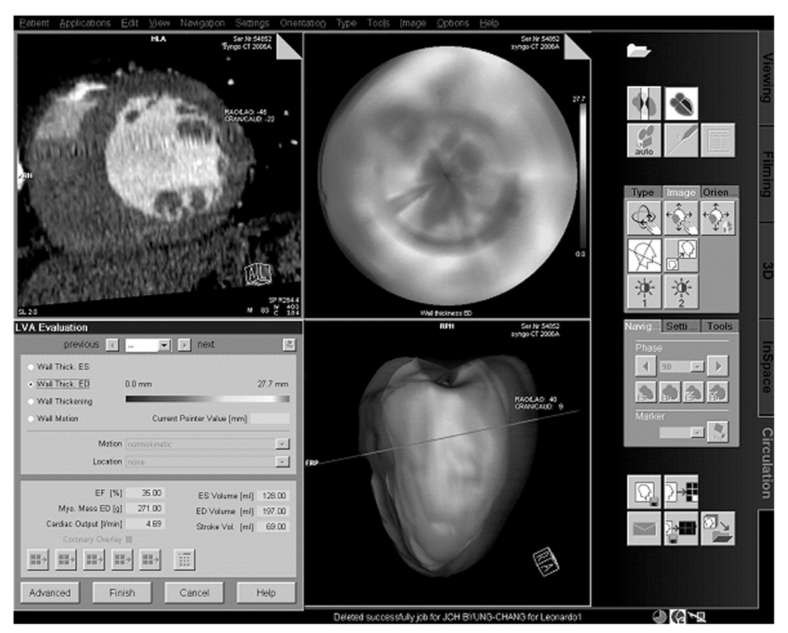
Figure 2
Short axis reformats from retrospectively ECG-gated MDCT allow circulation of end-systolic (A) and end-diastolic (B) images for assessment of ventricular function.
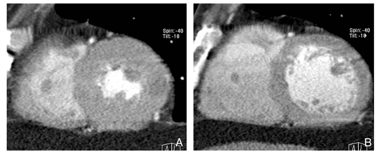
Figure 3
Images obtained in a 63-year-old man with acute myocardial infarction.
MR perfusion image (A) shows low signal area in the mid-inferior wall of left ventricle.
Delayed enhanced MR image (B) shows subendocardial dark zone (arrow) surrounded by hyperenhancement (arrowhead) in the same area.
Two-phase contrast enhanced MDCT shows early transmural perfusion defect (arrow) (C) and subendocardial residual perfusion defect (arrow) with subepicardial late enhancement (arrowhead) (D) in the mid inferior wall of left ventricle. This myocardial enhancement pattern correlates well with contrast-enhanced MR imaging
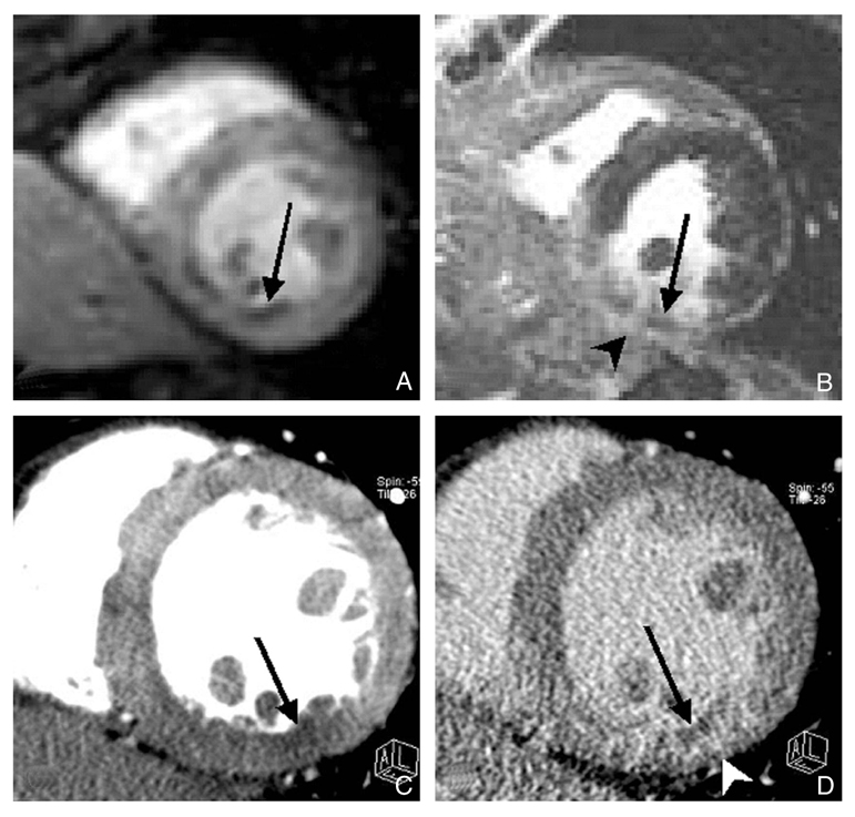
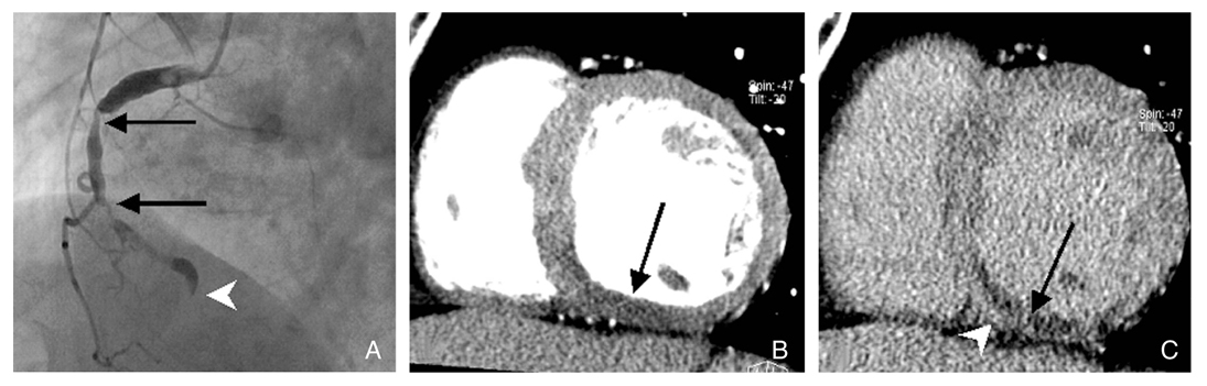
Figure 4
Images obtained in a 72-year-old man with reperfused acute myocardial infarction.
Catheter coronary angiogram (A) shows multiple significant stenoses in the proximal and middle (arrows) right coronary artery and total occlusion at the distal segment (arrowhead) of the right coronary artery. Two-phase contrast enhanced MDCT shows early transmural perfusion defect (B) and subendocardial residual perfusion defect (arrow) with subepicardial late enhancement (arrowhead) (C) in the mid-inferior wall of left ventricle.
- TOOLS
-
- Share :
-


-
METRICS

-
- 0 Crossref
- Scopus
- 1,396 View
- 5 Download
-

-
Related articles in
J Korean Med Assoc
- Editorial Office
-
37 Ichon-ro 46-gil, Yongsan-gu, Seoul
Tel: +82-2-6350-6562 Fax: +82-2-792-5208 E-mail: jkmamaster@gmail.com
Copyright © 2024 by Korean Medical Association.
