


 |
 |
| J Korean Med Assoc > Volume 50(1); 2007 > Article |
Abstract
The initial radiologic evaluation of a patient with acute abdominal symptoms begins with plain abdominal radiographs. Plain abdominal radiographs are helpful for the diagnosis of intestinal obstruction and pneumoperitoneum. However, cross-sectional imaging modalities, such as ultrasonography or computed tomography, are necessary for specific diagnosis of acute abdomen. Ultrasonography is a non-invasive and comfortable tool for patients visiting emergency room. This article describes the ultrasonographic findings of most common diseases presenting with acute abdominal symptoms.
References
1. Davies AH, Mastorakou I, Cobb R, Rogers C, Lindsell D, Mortensen NJM. Ultrasonography in the acute abdomen. Br J Surg 1991;78:1178-1180.
2. Laing FC. Ultrasonography of the acute abedomen. Radiol Clin North Am 1992;30:389-404.
3. Heller MB, Verdile VP. Ultrasonography in emergency medicine. Emerg Med Clin North Am 1992;10:27-46.
4. Hudson PA, Promes SB. Abdominal ultrasonography. Emerg Med Clin North Am 1997;15:825-848.
5. Cosgrove D, Meire H, Dewbury K. Abdominal and general ultrasound 1994;1st ed. Churchill Livingstone.
6. Lim JH, Kim PN, et al. Abdominal Radiology 2005;1st ed. Seoul: Korean Society of Abdominal Radiology.
7. O'Malley M, Wilson SR. US of gastrointestinal tract abnormalities with CT correlation. Radiographics 2003;23:59-72.
8. Vijayaraghavan SB. High-resolution sonographic spectrum of diverticulosis, diverticulitis, and their complications. J Ultrasound Med 2006;25:75-85.
9. Baker JA, Mandavia D, Swadron SP. Diagnosis of diverticulitis by bedside ultrasound in the emergency department. J Emerg Med 2006;30:327-329.
10. Singh AK, Gervais DA, Hahn P, Sagar P, Mueller PR, Novelline RA. Acute epiploic appendatitis and its mimics. Radiographics 2005;25:1521-1534.
11. Hollerweger A, Macheiner P, Rettenbacher T, Gritzmann N. Primary epiploic appendagitis: sonographic findings with CT correlation. Journal Clinical Ultrasound 2002;30:481-495.
12. Buljevac M, Busic Z, Cabrijan Z. Sonographic diagnosis of gallstone ileus. J Ultrasound Med 2004;23:1395-1398.
13. Lassandro F, Gagliardi N, Scuderi M, Pinto A, Gatta G, Mazzeo R. Gallstone ileus analysis of radiological findings in 27 patients. Eur J Radiol 2004;50:23-29.
14. Zubaidi A, Al-Saif F, Silverman R. Adult intussusception: A retrospective review. Dis Colon Rectum 2006;49:1-6.
15. Mateen MA, Saleem S, Rao PC, Gangadhar V, Reddy DN. Transient small bowel intussusception: ultrasound findings and clinical significance. Abdom Imaging 2006;30:1-7.
Figure 1
Normal appendix. Ultrasonogram using high frequency transducer on RLQ shows thin-walled, collapsed appendix (white arrows)
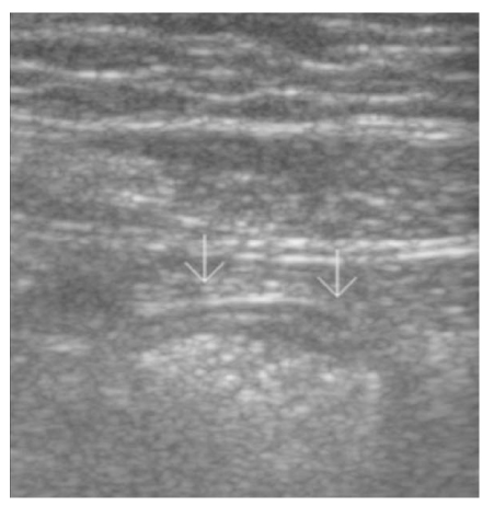
Figure 2
(A) Appendicitis. Longitudinal scan of ultrasonogram on RLQ shows thick-walled, distended appendix (diameter>6mm)
(B) Color Doppler ultrasonogram shows increase blood flow in inflamed appendix
(C) There is appendicolith in the tip of appendix on ultrasonogram
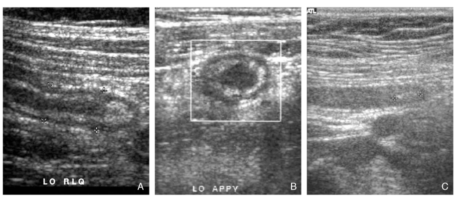
Figure 3
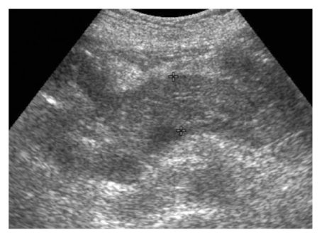
Acute cholecystitis. Longitudinal scan of RUQ shows large gallstone (white arrows) with wall thickening of gallbladder
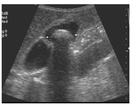
Figure 4
Diverticulitis in the ascending colon. Ultrasonogram using high frequency transducer (A) shows thickened wall of ascending colon (AC) and out-pouching sac with wall thickening (arrows). Computed tomography of lower abdomen (B) shows inflamed diverticulum (white arrow) and mesenteric infiltration (starlet)
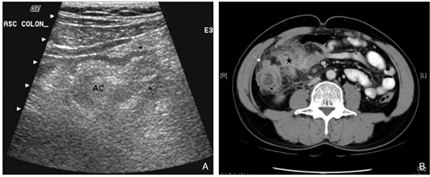
Figure 5
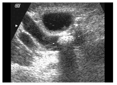
Appendagitis in the descending colon. Ultrasonogram in LLQ (A) shows well circumscribed echogenic mass with thin low echoic rim (white arrows) adjacent to descending colon. CT scan shows oval fatty mass (white arrows) with thin rim and internal high attenuation attached anterior wall of descending colon (B)
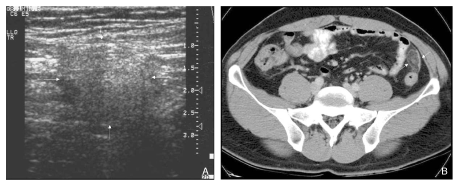
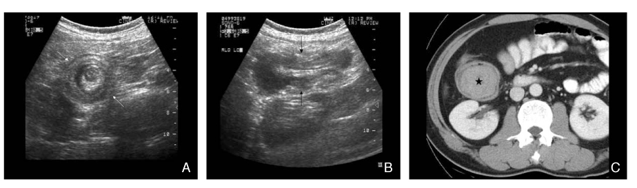
Figure 6
Gallstone ileus
(A) Ultrasonogram of lower abdomen shows large curvilinear bright echo (white arrows) with posterior echo shadowing in dilated small intestine
(B) This bright echo reveals to be stone (white arrows) on CT
(C) Ultrasonogram on RUQ shows collapse of GB and bright echoes in the lumen of gallbladder indicating airs (arrows)

Figure 7
Intussusception of small intestine. Transverse (A) and longitudinal (B) ultrasonograms of lower abdomen show multiple layered wall of small intestine with low echogic leading mass. CT scan (C) shows homogeneous enhancing mass (starlet) at the end of the intussusceptum, revealed to be B-cell lymphoma of small intestine
- TOOLS
-
- Share :
-


-
METRICS

-
- 1 Crossref
- Scopus
- 1,317 View
- 9 Download
-

-
Related articles in
J Korean Med Assoc -
Pornography and Sex in Adolescents1997 October;40(10)
Color Ultrasonography as Fetal Well-being Test1998 January;41(1)
- Editorial Office
-
37 Ichon-ro 46-gil, Yongsan-gu, Seoul
Tel: +82-2-6350-6562 Fax: +82-2-792-5208 E-mail: jkmamaster@gmail.com
Copyright © 2024 by Korean Medical Association.
