


 |
 |
| J Korean Med Assoc > Volume 49(8); 2006 > Article |
Abstract
High intensity focused ultrasound (HIFU) is a technique that was first introduced in the 1940s as a potential method of destroying selective regions within the brain to aid neurobehavioral studies. A beam of ultrasound can be delivered to a targeted focus at a distance from its source, and if a sufficient amount of energy is concentrated in the focus, the cells lying within this focal volume are selectively killed. This is, therefore, a non-invasive method of producing selective and "trackless" tissue destruction in deep-seated targets in the body without damage to the overlying tissues. Although it had not been in clinical use for a long time, HIFU is now widely used as a non-invasive treatment method for malignant tumors of the liver, kidney, breast, bone, uterus and pancreas, as well as for the relief of chronic pain of malignant origin. Further improvement of technology and imaging of HIFU in the near future will make it one of the most important tools in the treatment of solid tumors, further expanding its clinical applications.
References
1. Vogl TJ, Straub R, Eichler K, Soller O, Mack MG. Colorectal carcinoma metastases in liver: laser-induced interstitial thermotherapy : local tumor control rate and survival data. Radiology 2003;230:450-458.
2. Nagaoka Y, Nakayama R, Iwata M. Cutaneous seeding following percutaneous ethanol injection therapy for hepatocellular carcinoma. Intern Med 2004;43:268-269.
3. Liu C, Frilling A, Dereskewitz C, Broelsch CE. Tumor seeding after fine needle aspiration biopsy and percutaneous radiofrequency thermal ablation of hepatocellular carcinoma. Dig Surg 2003;20:460-463.
4. Thuroff S, Chaussy C, Vallancien G, Wieland W, Kiel HJ, Gelet A, et al. High-intensity focused ultrasound and localized prostate cancer : efficacy results from the European multicentric study. J Endourol 2003;17:673-677.
5. Kennedy JE, Wu F, ter Haar GR, Gleeson FV, Phillips RR, Middleton MR, et al. High-intensity focused ultrasound for the treatment of liver tumours. Ultrasonics 2004;42:931-935.
6. Wu F, Wang ZB, Chen WZ, Zou JZ, Bai J, Gao GW, et al. Extracorporeal focused ultrasound surgery for treatment of human solid carcinomas: early Chinese clinical experience. Ultrasound Med Biol 2004;30:245-250.
7. Gianfelice D, Kheat A, Boulanger Y, Amara M, Belblidia A. Feasibility of magnetic resonance imaging-guided focused ultrasound surgery as an adjunct to tamoxifen therapy in high-risk surgical patients with breast carcinoma. J Vasc Interv Radiol 2003;14:1275-1282.
8. Wu F, Wang ZB, Chen WZ, Bai J, Zhu H, Qiao TY. Preliminary experience using high intensity focused ultrasound for the treatment of patients with advanced stage renal malignancy. J Urol 2003;170:2237-2240.
9. Chen L, Rivens L, ter Haar G, Riddler S, Hill CR, Bensted JP. Histological changes in rat liver tumours treated with high-intensity focused ultrasound. Ultrasound Med Biol 1993;19:67-74.
10. ter Haar G, Clarke RL, Vaughan MG, Hill CR. Trackless surgery using focused ultrasound: technique and case report. Minimally invasive Therapy 1991;1:13-19.
11. Hill CR, ter Haar GR. High intensity focused ultrasound: potential for cancer treatment. Br J Radiol 1995;68:1296-1303.
12. Wu F, Chen WZ, Bai j, Zou JZ, Wang ZL, Wang ZB, et al. Pathological changes in human malignant carcinoma treated with high-intensity focused ultrasound. Ultrasound Med Biol 2001;27:1099-1106.
13. Wu F, Wang ZB, Jin CB, Zhang JP, Chen WZ, Zou JZ, et al. Circulating tumor cells in patients with solid malignancy treated by high-intensity focused ultrasound. Ultrasound Med Biol 2004;30:511-517.
14. Vallejo R, Hord ED, Barna SA, Santiago-Palma J, Ahmed S. Perioperative immunosuppression in cancer patients. J Environ Pathol Toxicol Oncol 2003;22:139-146.
15. Mafune K, Tanaka Y. Influence of multimodality therapy on the cellular immunity of patients with esophageal cancer. Ann Surg Oncol 2000;7:609-616.
16. Kramer G, Steiner GE, Grobl M, Hrachowitz K, Reithmayr F, Marberger M, et al. Response to sublethal heat treatment of prostatic tumor cells and of prostatic tumor infiltrating T-cells. Prostate 2004;58:109-120.
17. Visioli AG, Rivens IH, ter Haar GR, Horwich A, Huddart RA, Glees J, et al. Preliminary results of a phase I dose escalation clinical trial using focused ultrasound in the treatment of localized tumours. Eur J Ultrasound 1999;9:11-18.
18. Vallancien G, Harouni M, Guillonneau B, Veillon B, Bougaran J. Ablation of superficial bladder tumors with focused extracorporeal pyrotherapy. Urology 1996;47:204-207.
19. Kennedy JE. High-intensity focused ultrasound in the treatment of solid tumors. Cancer 2005;5:321-327.
20. Fry WJ, Mosberg WH, Bamard JW, Fry FJ. Production of focal destructive lesions in the central nervous system with ultrasound. J Neurosurg 1954;11:471-478.
21. Blana A, Walter B, Rogenhofer S, Wieland WF. High-intensity focused ultrasound for the treatment of localized prostate cancer: 5-year experience. Urology 2004;63:297-300.
22. Gelet A, Chapelon JY, Poissonnier L, Bouvier R, Rouviere O, Vallancien G, et al. Local recurrence of prostate cancer after external beam radiotherapy: early experience of salvage therapy using high-intensity focused ultrasonography. Urology 2004;63:625-629.
23. Stewart EA, Gedroyc WM, Tempany CM, Quade BJ, Inbar Y, Rabinovici J, et al. Focused ultrasound treatment of uterine fibroid tumors: safety and feasibility of a noninvasive thermoablative technique. Am J Obstet Gynecol 2003;189:48-54.
24. McDannold N, Moss M, Killiany R, Rosene DL, King RL, Hynynen K, et al. MRI-guided focused ultrasound surgery in the brain: tests in a primate model. Magn Reson Med 2003;49:1188-1191.
25. Wu F, Wang ZB, Cao YD, Chen WZ, Bai J, Zhu H, et al. A randomised clinical trial of high-intensity focused ultrasound ablation for the treatment of patients with localised breast cancer. Br J Cancer 2003;89:2227-2233.
26. Marbeger M, Schatzl G, Cranston D, Kennedy JE. Extracorporeal ablation of renal tumors with high intensity focused ultrasound. Br J Urol 2005;95:Suppl 2. 52-55.
27. Wu F, Wang JB, Chen WZ, Zou JZ, Bai J, Su HB, et al. High intensity focused ultrasound ablation combined with transcatheter arterial embolisation in the treatment of advanced hepatocellular carcinoma. Radiology 2005;235:659-667.
28. Bohris C, Jenne JW, Rastert R, Simiantonakis I, Brix G, Debus J, et al. MR monitoring of focused ultrasound surgery in a breast tissue model in vivo. Magn Reson Imaging 2001;19:167-175.
29. Sedelaar JP, Aarnink RG, van Leenders GJ, Beerlage HP, Debruyne FM, de La Rosette JJ, et al. The application of three-dimensional contrast-enhanced ultrasound to measure volume of affected tissue after HIFU treatment for localized prostate cancer. Eur Urol 2000;37:559-568.
30. Anderson GS, Brinkmann F, Soulen MC, Alavi A, Zhuang H. FDG positron emission tomography in the surveillance of hepatic tumors treated with radiofrequency ablation. Clin Nucl Med 2003;28:192-197.
31. Cannon JW, Stroll JA, Salgo IS, Knowles HB, Howe RD, del Nido PJ, et al. Real-time three-dimensional ultrasound for guiding surgical tasks. Comput Aided Surg 2003;8:82-90.
32. Righetti R, Kallel F, Stafford RJ, Price RE, Krouskop TA, Ophir J, et al. Elastographic characterization of HIFU-induced lesions in canine livers. Ultrasound Med Biol 1999;25:1099-1113.
33. Penney GP, Blackall JM, Hamady MS, Sabharwal T, Adam A, Hawkes DJ. Registration of freehand 3D ultrasound and magnetic resonance liver images. Med Image Anal 2004;8:81-91.
Figure 1
Schematic showing the principle of high-intensity focused ultrasound
A) An extracorporeal source generates an ultrasound beam, which forms a cigar-shaped focus deep within the target tissue(liver). The volume of ablation('lesion') following a single high-intensity focused ultrasound exposure is small and will vary according to transducer characteristics, but is typically in the order of 1~3mm wide by 8~15mm in length along the beam axis.
B) Schematic illustrating application of sequential "single lesions" to achieve tumour volume ablation. The lesions must be placed side by side systematically to "paint out" the target tumour and some of the surrounding normal tissue margin.

Figure 2
The principle of inertial cavitation
A mechanical ultrasound wave progresses through tissues(top), causing alternating cycles of increased and reduced pressure (compression and rarefaction respectively-middle). Gas is drawn out of solution during rarefaction, creating bubbles. These can oscillate in size in a stable fashion with the chaging tissue pressure, but ultimately might collapse, causing local energy release and temperature rises at the microscopic level (bottom).
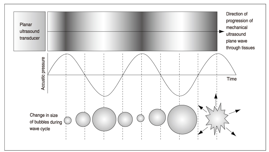
Figure 3
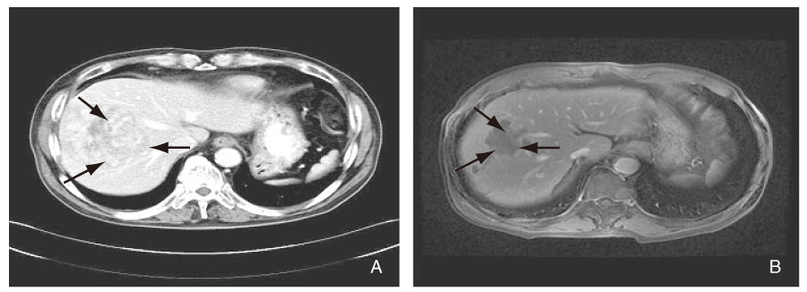
A case of high-intensity focused ultrasound for hepatocelluar carcinoma
A) Before HIFU: Abdominal CT shows a large, 9cm-diameter, hepatocelluar carcinoma(arrows) at right hepatic lobe in 67-year-old man.
B) After HIFU: MR obtained 2 months later shows the lesion(arrows) has markedly decreased without contrast enhancement.
- TOOLS
-
- Share :
-


-
METRICS

-
- 1 Crossref
- Scopus
- 1,104 View
- 2 Download
-

-
Related articles in
J Korean Med Assoc
- Editorial Office
-
37 Ichon-ro 46-gil, Yongsan-gu, Seoul
Tel: +82-2-6350-6562 Fax: +82-2-792-5208 E-mail: jkmamaster@gmail.com
Copyright © 2024 by Korean Medical Association.
