


 |
 |
| J Korean Med Assoc > Volume 47(9); 2004 > Article |
Abstract
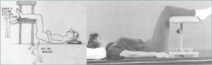
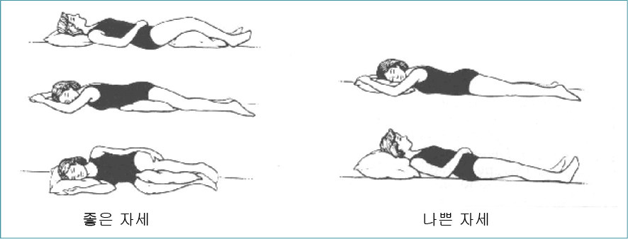
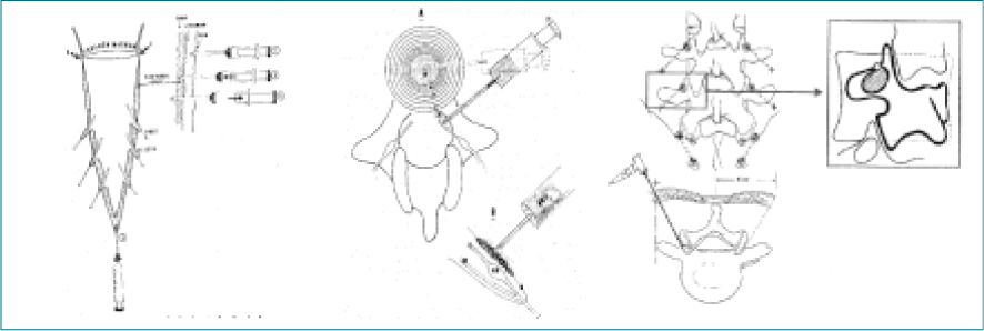
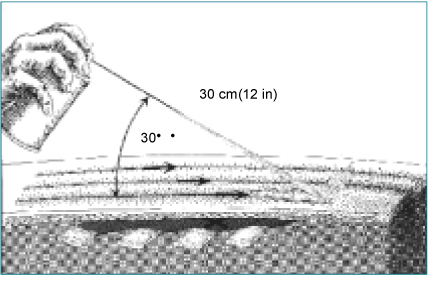
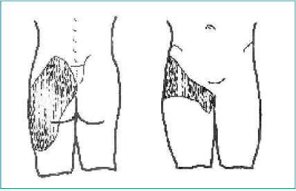
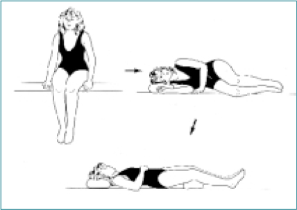
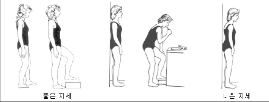
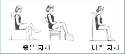
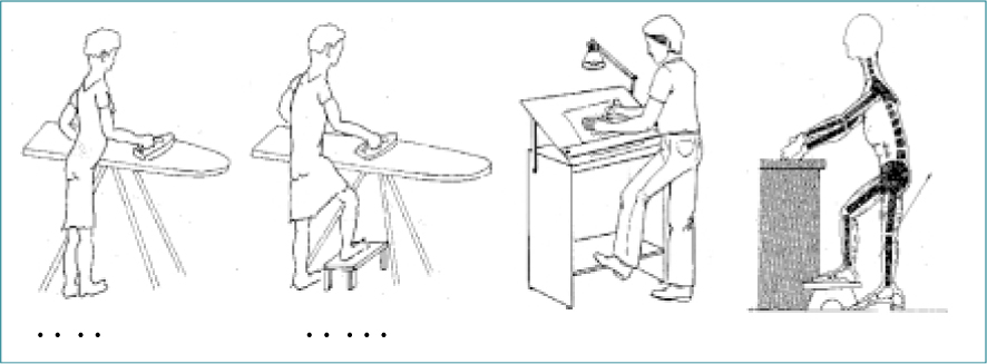
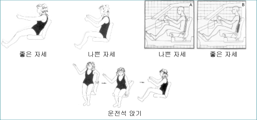
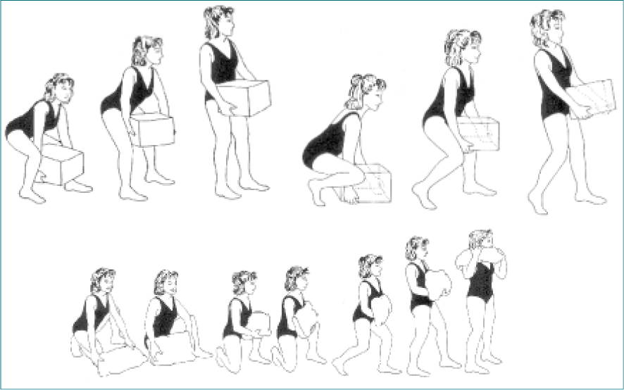
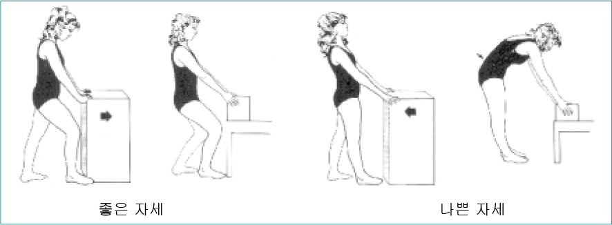
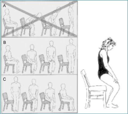
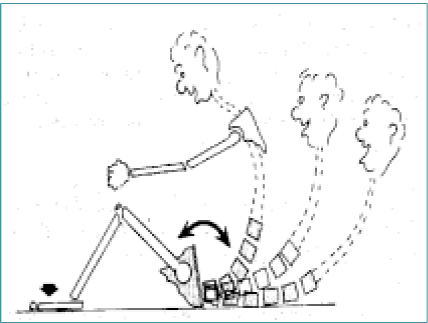
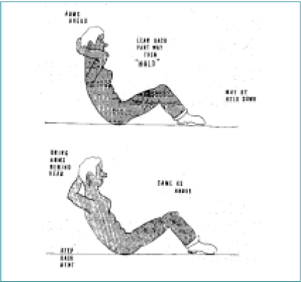
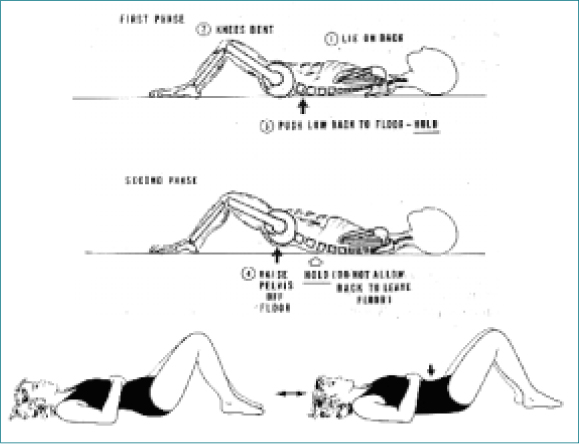
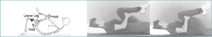
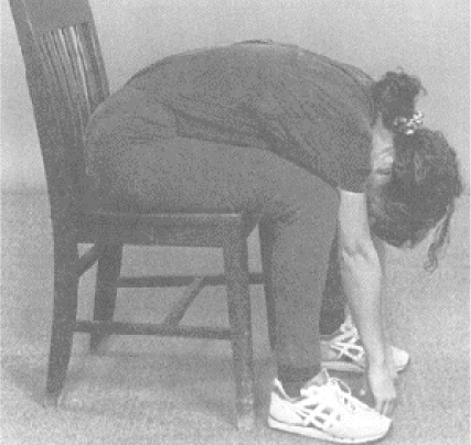
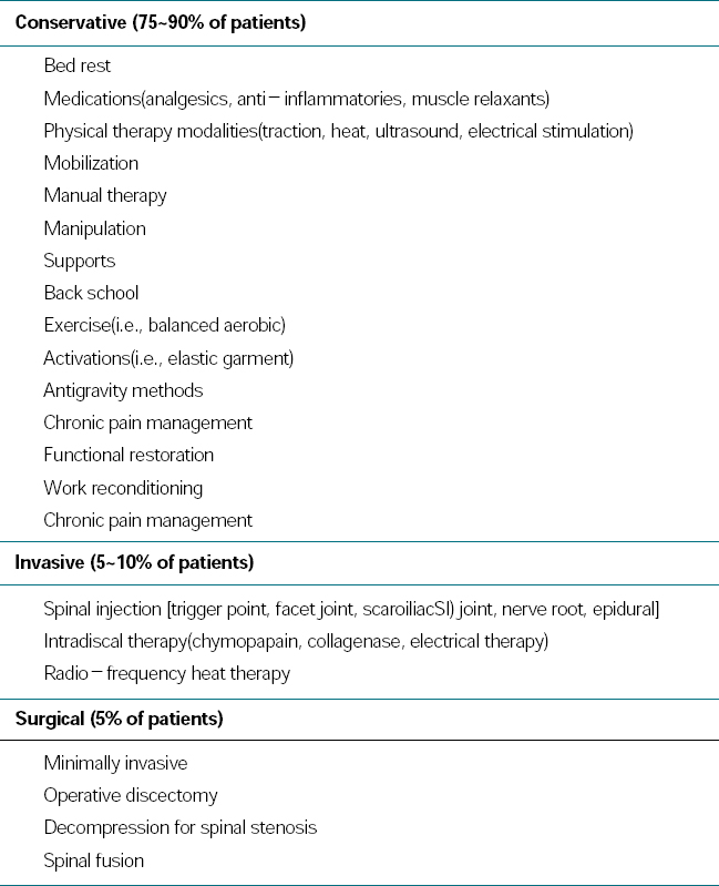
Most people will experience episodes are usually brief, resolve spontaneously, and recur infrequently. The successful management of persistent low back pain requires that treatment be directed to the pain-producing structures in the human body. The spectrum of the treatment of low back pain ranges from very simple and straight foreword to very the complex and intricate. Treatments for lumbar disc herniations are conservative (75~90% of patients), invasive (5~10% of patients), and surgical (5% of patients) treatments. Resolution of the first lumbar disc herniation takes place in approximately 75% of patient over a period of 3 months. With recurrent herniations, the chance of spontaneous relief of symptoms is reduced. In a very acute stage, the patient may require hospitalization to control the level of pain. Bed rest should be limited 2 days with the most comfortable position of the knee and the hip flexion about 80~90 degree. A few days of bed rest, adequate analgesics, and muscle relaxants to reduce muscle spasm usually are require. Physical therapeutic modality(included traction, heat, ultrasound, electrical stimulation), mobilization, manipulation, back school, spinal supports, therapeutic exercise and proper position should be used and educated. If the patient did not controled low back pain after above treatments, invasive treatments such as trigger point injection, facet or sacroiliac joint injection, epidural steroid injection, selective nerve root injection with high frequency heat therapy, or intradiscal injection may quickly alleviate symptoms. Every patient should attend a class in spine education as part of the overall treatment. Instruction is given in low back care, especially as related to the activities of daily living. Participants are taught correct posture, pelvic tilting, knee-to-chest exercise, and exercises to strengthen abdominal and paraspinal muscles. Individual instructions are given to each patient, explaining in more detail the nature of the patient's particular problem and how the individual can take control of the treatment.
References
5. Arokoski JP, Valta T, Airaksinen O, Kankaanpaa M. Back & abdominal muscle function during stabilization exercises. Arch Phys Med Rehabil 2001;82:1089-1098.
6. Braddom RL. Physical Medicine & Rehabilitation 2000;2nd ed. Philadelphia: WB Saunders.
7. Cailliet R. Pain series : Low back pain syndrome 1995;5th ed. F.A. Davis Company.
8. Cox JM. Low back pain : mechanism, diagnosis and treatment 1999;6th ed. Boltimore: Williams & Wlikins.
9. Delisa JA, Gans BM. Rehabilitation Medicine, principles and practice 1998;3rd ed. Philadelphia: Lippincott Company.
10. Kirkaldy-Willis WH, Burton CV. . Managing low back pain 1992;4th ed. London: Churchill Livingstone.
11. Leinonen V, Kankaanpää , Luukkonen M, Hanninen O, Airaksinen O, Taimela S. Disc herniation-related back pain impairs feed-forward control of paraspinal muscles. Spine 2001;26:E367-E372.
12. Mannion AF, Taimela S, Muntener M, Dvorak J. Active therapy for chronic low back pain : part 1. Effects on back muscle activation, fatigability, and strength. Spine 2001;26:897-908.
13. Richardson C, Jull G, Hodges P, Hides J. Therapeutic exercises for spinal segmental stabilization in low back pain 1999;London: Churchill Livingstone.
14. Travell JG, Simons DG. Myofascial Pain and Dysfunction 2000;illustrator: Barbara D. Cummings.
15. van Tulder MW, Koes BW, Bouter LM. Conservative treatment of acute and chronic nonspecific low back pain : a systematic review of randomized controlled trials of the most common interventions. Spine 1997;22:2128-2156.
16. van Tulder MW, Malmivaara A, Esmail R, Koes BW. Exercise therapy for low back pain. Cochrane Database Syst Rev 2000.
- TOOLS
-
- Share :
-


-
METRICS

-
- 1 Crossref
- Scopus
- 1,089 View
- 14 Download
-

-
Related articles in
J Korean Med Assoc -
Pain Management in Cancer Patients2001 December;44(12)
Surgical Treatment of Degenerative Lumbar Disc Disease2004 September;47(9)
Physical Therapy and Pharmacological Treatment of Lumbar Disc Herniations2004 September;47(9)
Current Management of Peripheral Arterial Disease2010 March;53(3)
Requirements and circumstances for safe sedation2013 April;56(4)
- Editorial Office
-
37 Ichon-ro 46-gil, Yongsan-gu, Seoul
Tel: +82-2-6350-6562 Fax: +82-2-792-5208 E-mail: jkmamaster@gmail.com
Copyright © 2024 by Korean Medical Association.
