16-MDCT : A New Modality for Diagnosis of Cardiac Diseases

Article information

J Korean Med Assoc. 2003;46(6):535-541
Publication date (electronic) : 2003 June 30
doi : https://doi.org/10.5124/jkma.2003.46.6.535
Department of Diagnostic Radiology, University of Ulsan College of Medicine, Asan Medical Center, Korea. thlim@amc.seoul.kr

Abstract

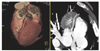
Since the advent of 16-MDCT in the clinical diagnosis, a paradigm shift is required in the diagnostic algorithm of cardiovascular diseases owing to its revolutionary technical advances. 16-MDCT provides less than 1 mm of high spatial resolution even in z-direction by using detectors with less than 0.75 mm collimation, which in turn allows for volumetric scanning with isotropic 3-dimensional resolution. This high spatial resolution of 16-MDCT enables one to obtain high quality images of the small vessels such as coronary arteries with diameter < 1 mm. For imaging of the heart, scanning with a high temporal resolution is particularly important because of the strong movement during the cardiac cycle. 16-MDCT allows images with a high temporal resolution, less than 250 msec, enough to freeze the cardiac motion. Furthermore, by using ECG information that is recorded simultaneouly with the image acquisition, images synchronized to the specific cardiac phase can either be scanned or post-processed according to the technique of ECG-gating. In order to eliminate the motion artifact from respiratory motion, scanning must be completed within a single breathholding time. By adopting 12~16 detector arrays and less than 0.5 sec of gantry rotation time, imaging of the whole heart with submillimeter spatial resolution can be covered within 20 seconds of breathholding time. Major clinical applications of 16-MDCT in cardiac diseases inlcude detection of coronary stenosis and atherosclerotic plaque, coronary calcium scoring, evaluation of the patients after coronary angioplasty or coronary arterial by-pass graft, assessment of the cardiac valve morphology and function, and ventricular function and perfusion. Among these, currently the most practical area of 16-MDCT application is post-CABG evaluation and the most imporatnt and promising area will be assessment of native coronary arteries for detection of stenotic vessels and for detecting and differentiating atherosclerotic lesions over a spectrum of vulnerable, soft, fibrotic, and calcified plaques. In this review, important technical aspects together with clinical applications of 16-MDCT in diagnosis of cardiovascular diseases will be presented.

References

1. Stehling MK, Turner R, Mansfield P. Echo-planar imaging magnetic resonance imaging in a fraction of a second. Science 1991. 25443–50.
2. Ohnesorge B, Flohr T, et al, eds. Technical aspects and applications of fast multislice cardiac CT. Medical radiology, diagnostic imaging and radiation oncology, multislice CT 2001. 1st edth ed. Berlin, Heidelberg, New York: Springer; 121–130.
3. Flohr T, Ohnesorge B. Heart-rate adaptive optimization of spatial and temporal resolution for ECG-gated multi-slice spiral CT of the heart. J Comput Assist Tomogr 2001. 25(6)907–923.
4. Hong C, Becker CR, Huber A, Schpf UJ, Ohnesorge B, Reiser MF, et al. ECG-gated reconstructed multi-detector row CT coronary angiography : effect of varying trigger delay on image quality. Radiology 2001. 220712–717.
5. Kopp AF, Schrder S, Kttner A, Heuschmid M, Georg C, Ohnesorge B, et al. Coronary arteries : retrospectively ECG-gated multi-detector row CT angiography with selective optimization of the reconstruction window. Radiology 2001. 221683–688.
6. Ropers D, Baum U, Pohle K, Anders K, Ulzheimer S, Ohnesorge B, et al. Detection of coronary artery stenoses with thin-slice multi-detector row spiral computed tomography and multiplanar reconstruction. Circulation 2003. 107664–666.
7. Nieman K, Cademartiri F, Lemos PA, Raaijmakers R, Pattynama PMT, de Feyter PJ. Reliable noninvasive coronary angiography with fast mubmillimeter multislice spiral computed tomography. Circulation 2002. 1062051–2054.
8. Kopp AF, Ohnesorge B, Becker C, Schrder S, Heuschmid M, Kttner A, et al. Reproducibility and accuracy of coronary calcium measurements with multi-detector row versus electron-beam CT. Radiology 2002. 225113–119.
9. O'Rourke RA, Brundage BH, Froelicher V, Greenland P, Grundy SM, Wolk MJ, et al. American College of Cardiology/American Hear Association expert consensus document on electron-beam computed tomography for the diagnosis and prognosis of coronary artery disease. Circulation 2000. 102126–140.
10. Hunold P, Vogt FM, Schmermund A, Debatin JF, Kerkhoff G, Barkhausen J, et al. Radiation exposure during cardiac CT : Effective doses at multi-detector row CT and electron-beam CT. Radiology 2003. 226145–152.
11. Yang M, Akbaki H, Reddy GP, Higgins CB. Identification of pulmonary vein stenosis after radiofrequency ablation for atrial fibrillation using MRI. J Comput Assist Tomogr 2001. 2534–35.
12. Willmann JK, Weishaupt D, Lachat M, et al. Electrocardiographically gated multi-detector row CT for assessement of valvular morphology and calcification in aortic stenosis. Radiology 2002. 225120–128.
13. Willmann JK, Kobza R, Roos JE, et al. ECG-gated multidetector row CT for assessment of mitral valve disease : initial experience. Eur Radiol 2002. 122662–2669.
14. Halliburton S, Petersilka M, Schvartzman P, et al. Validation of left ventricular volume and ejection fraction measurement with multi-slice computed tomography : comparison to cine magnetic resonance imaging. Radiology 2001. 221(P)452.
15. Mochizuki T, Higashino H, Kayama Y, et al. Evaluation of wall motion using multi-detector-row CT : new application of post-processing interactive multi-planar animation of the heart. Radiology 2001. 221(P)413.

Article information Continued

Figure 1

Figure 2

Figure 3

Figure 4

Figure 5