Status of common parasitic diseases in Korea in 2019
Article information
Abstract
Abstract
This study aimed to determine the status of common parasitic disease in Korea in 2019. Twelve parasitic diseases were selected: toxocariasis, anisakiasis, paragonimiasis, sparganosis, cysticercosis, toxoplasmosis, clonorchiasis, enterobiasis, trichuriasis, trichomoniasis, cryptosporidiosis, and malaria. Their biology, epidemiology, pathogenesis, symptoms and signs, diagnosis, treatment, and prognosis were evaluated. Of the parasitic diseases, toxocariasis was the most prevalent according to serological results. Anisakiasis should be considered when acute gastrointestinal symptoms occur with a recent past history of raw seafood ingestion. Paragonimiasis, sparganosis, and cysticercosis can be diagnosed using an enzyme-linked immunosorbent assay; thus, enzyme-linked immunosorbent assay needs to be performed for suspected cases. Toxoplasmosis and cryptosporidiosis are opportunistic infections. The symptoms and signs are aggravated under immunocompromised conditions. Although the egg positivity rate of Clonorchis sinensis is higher than that of other intestinal parasitic diseases, encountering patients with complaints of symptoms caused by clonorchiasis is rare because the worm burden is low. Trichomoniasis is usually managed by gynecologists; therefore, it should be included in the differential diagnoses of vaginal diseases. The annual number of malaria cases has decreased, although it remains at approximately 500 cases per year. Malaria should be suspected when symptoms such as intermittent fever, headache, and splenomegaly are noted especially when the patients reside near demilitarized zones. Although the prevalence and number of reported cases of parasitic diseases have decreased in Korea, we should consider parasitic diseases in the list of differential diagnoses.
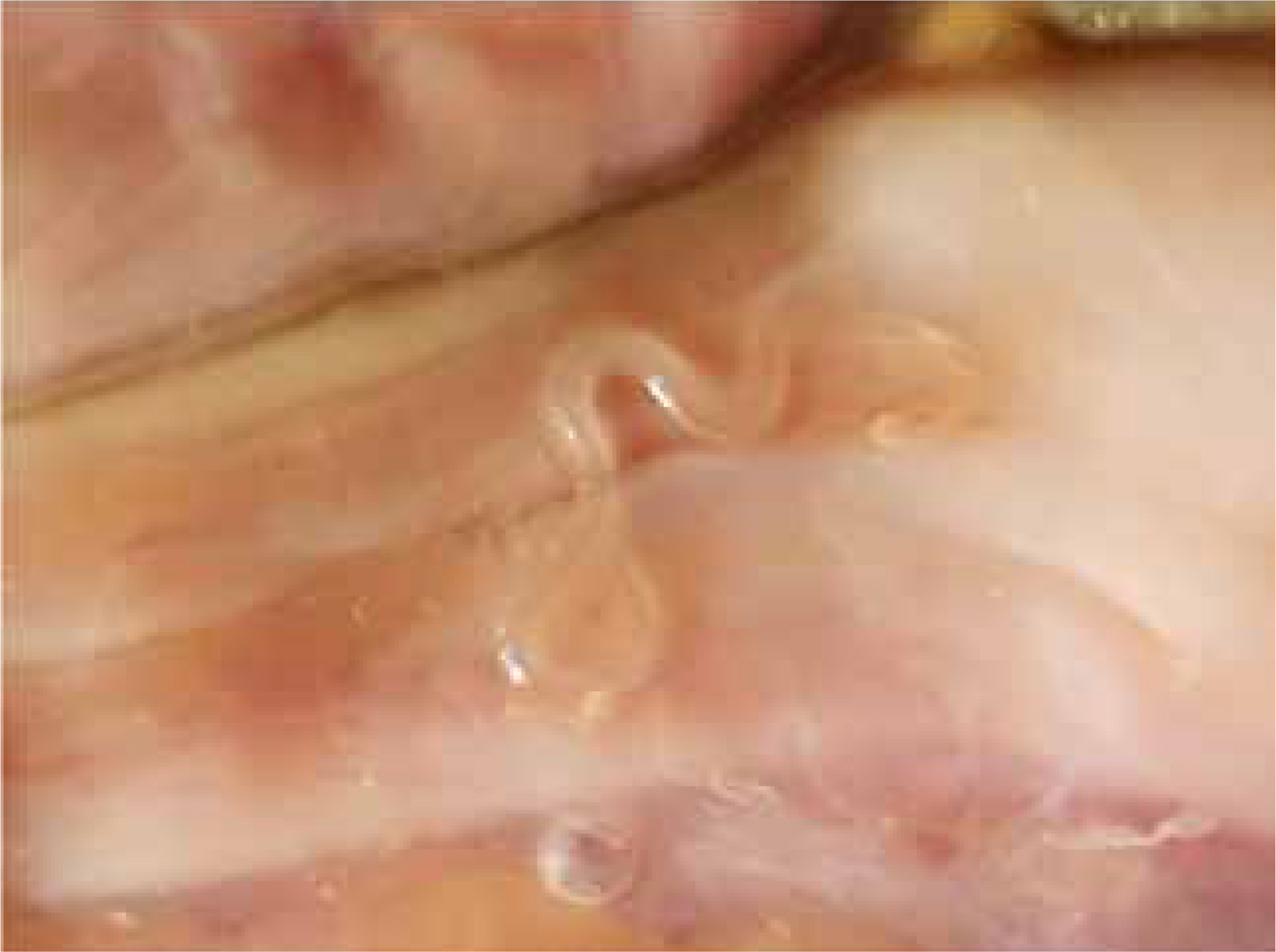
Anisakis larva in mesentry of Anago anago (photo by Sun Huh, 2019).
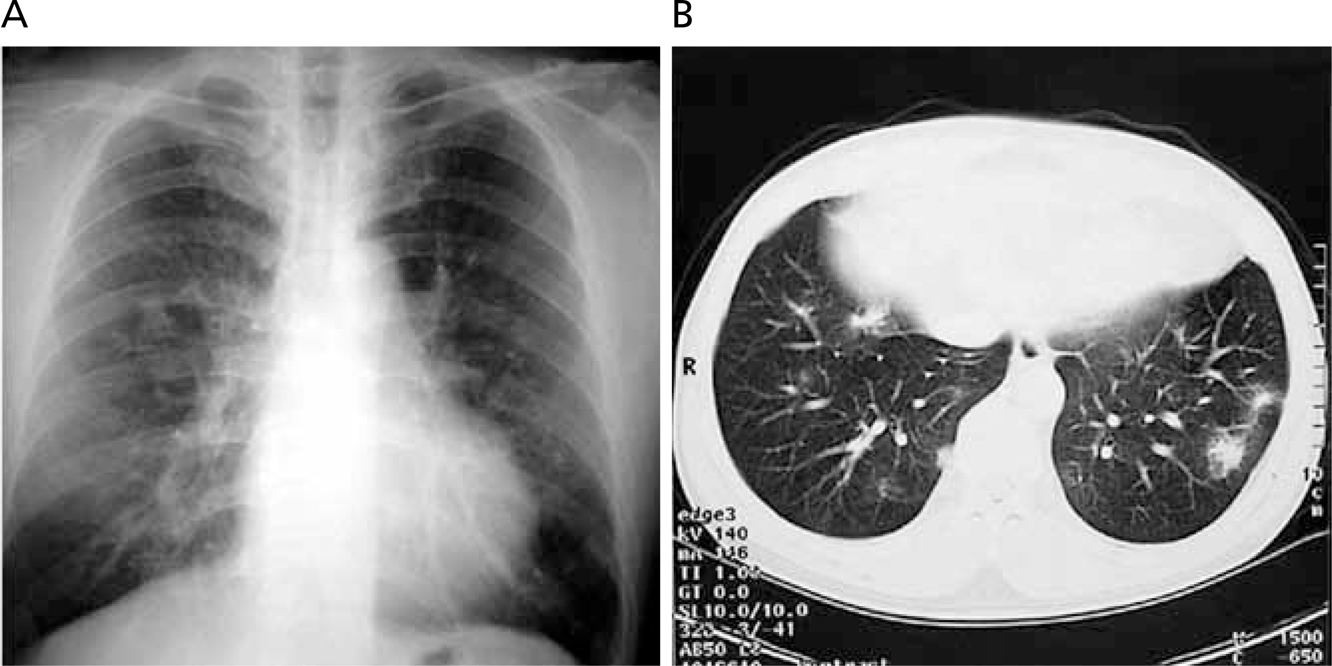
(A) Chest posteroanterior radiograph in a patient with toxocariasis. (B) A computed tomography scan of the patient with toxocariasis showing multiple pulmonary nodules with surrounding ground-glass opacities at first visit (Unpublished case).
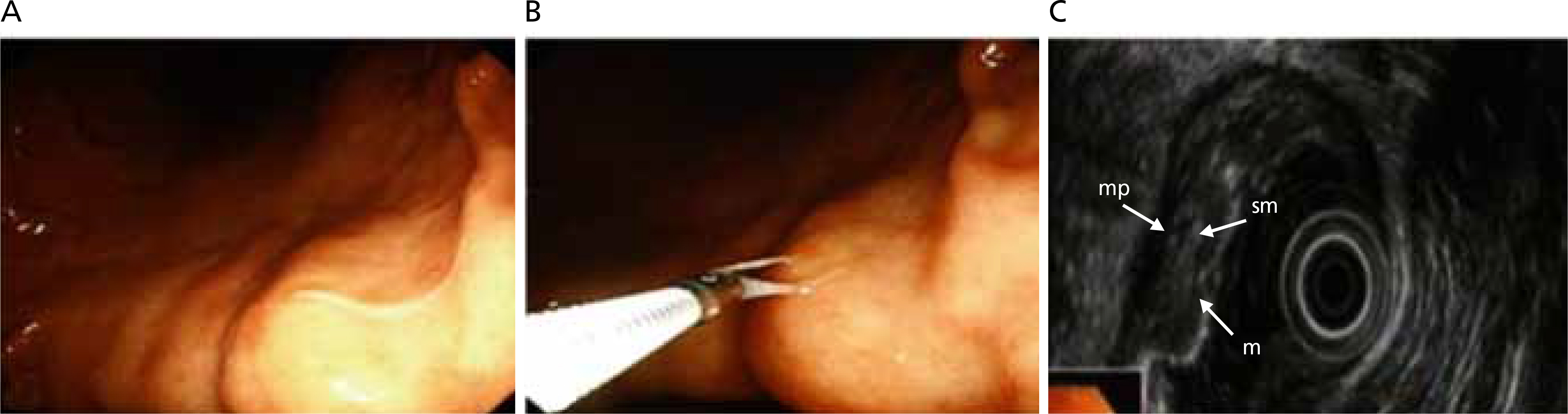
Endoscopic and endosonographic findings of the stomach. (A) An anisakid larva is penetrating into the mucosa of the lesser curvature of the gastric body. The gastric mucosa around the worm shows marked hyperemia. (B) The worm is captured by endoscopic forceps for removal. (C) On endosonography, gastric wall thickening can be seen. The most thickened layer is the submucosa (third layer of the stomach). Its character is homogenous and relatively hypodense. The 5-layered wall structure is well preserved. mp, muscularis propria; sm, submucosal layer; m, mucosal layer. Reproduced from Kim SH et al. Clin Endosc 2013;46:293-296, according to the Creative Commons license [14].
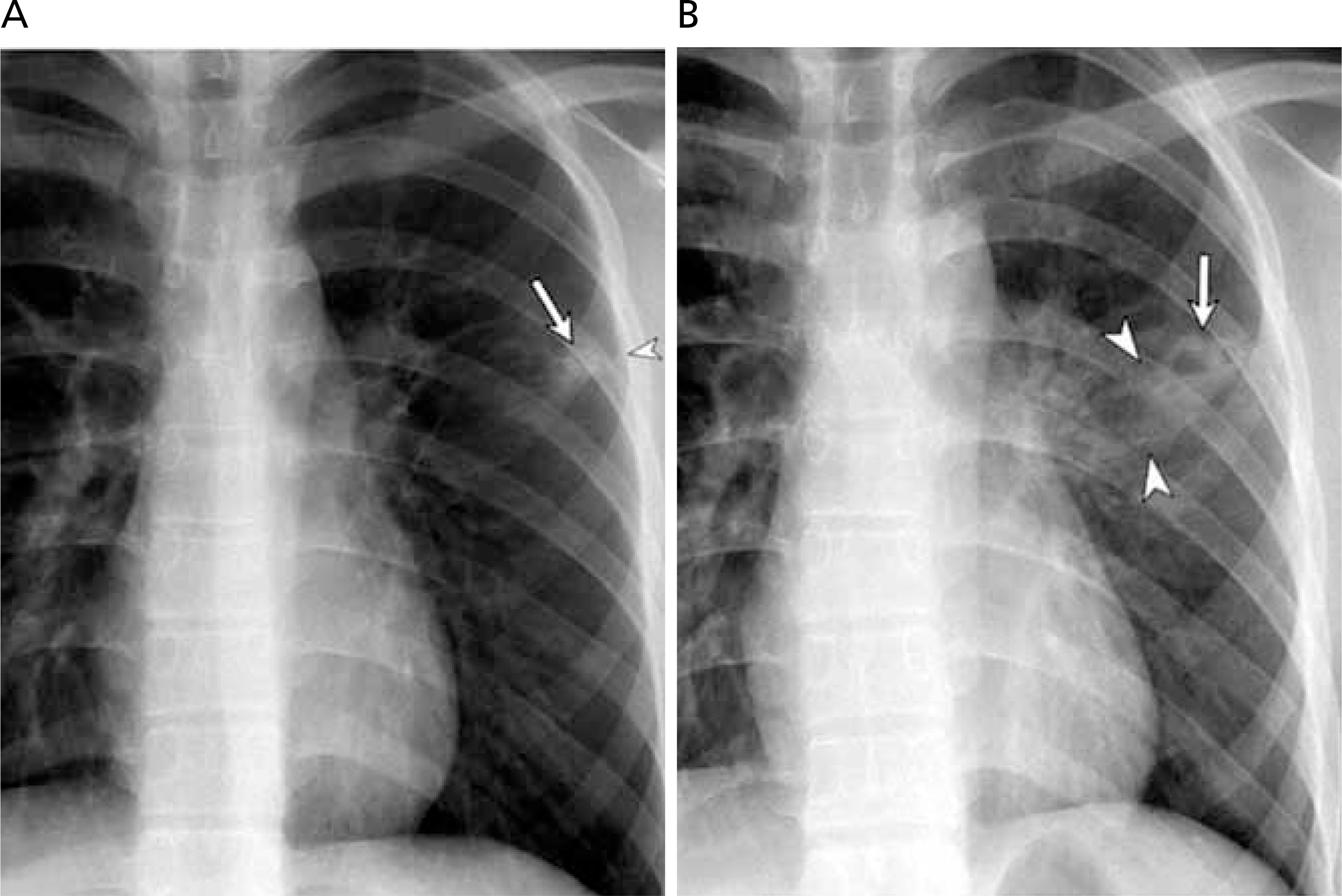
(A) Initial chest radiograph shows a subpleural nodule (arrow) in the left upper lung field with localized pleural thickening (arrowhead). Note subtle bubble-like air density within the nodule. (B) Follow-up chest radiograph after completion of antituberculosis medication reveals enlargement of the nodule with increased cavitary portion (arrow) accompanied with pulmonic infiltrates (arrowheads) in paragonimiasis. Reproduced from Park SE et al. Pediatr Infect Vaccine 2017;24:178-182, according to the Creative Commons license [17].

Intraoperative findings of sparganosis case. (A) A lesion is located between the left upper outer part of the breast and the axilla. (B) Skin incision is done over the main lesion and a worm is detected between the pectoralis muscles. (C) A close-up picture of the worm. (D) A worm is being extracted. Reproduced from Oh MY et al. Korean J Parasitol 2019;57:179-184, according to the Creative Commons license [23].
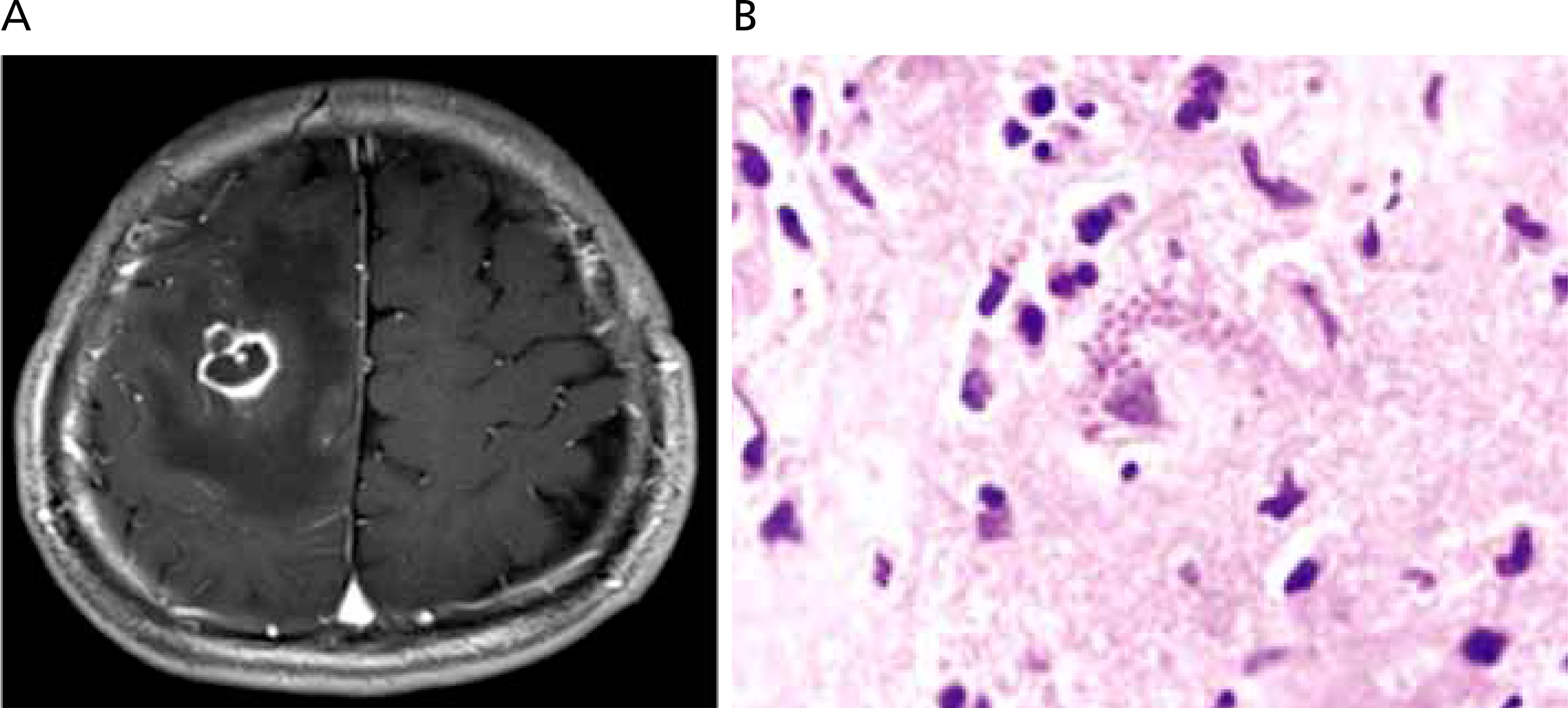
(A) Magnetic resonance imaging showing a ring-enhanced mass with mild edema in the right frontal lobe. (B) Many minute, basophilic bradyzoites fill a ruptured protozoal pseudocyst of Toxoplasm gondii with surrounding brain tissue which shows edema and infiltrating inflammatory cells (hematoxylin-eosin stain, ×200). Reproduced from Lee SB et al. Brain Tumor Res Treat 2017;5:34-36, according to the Creative Commons license [28].
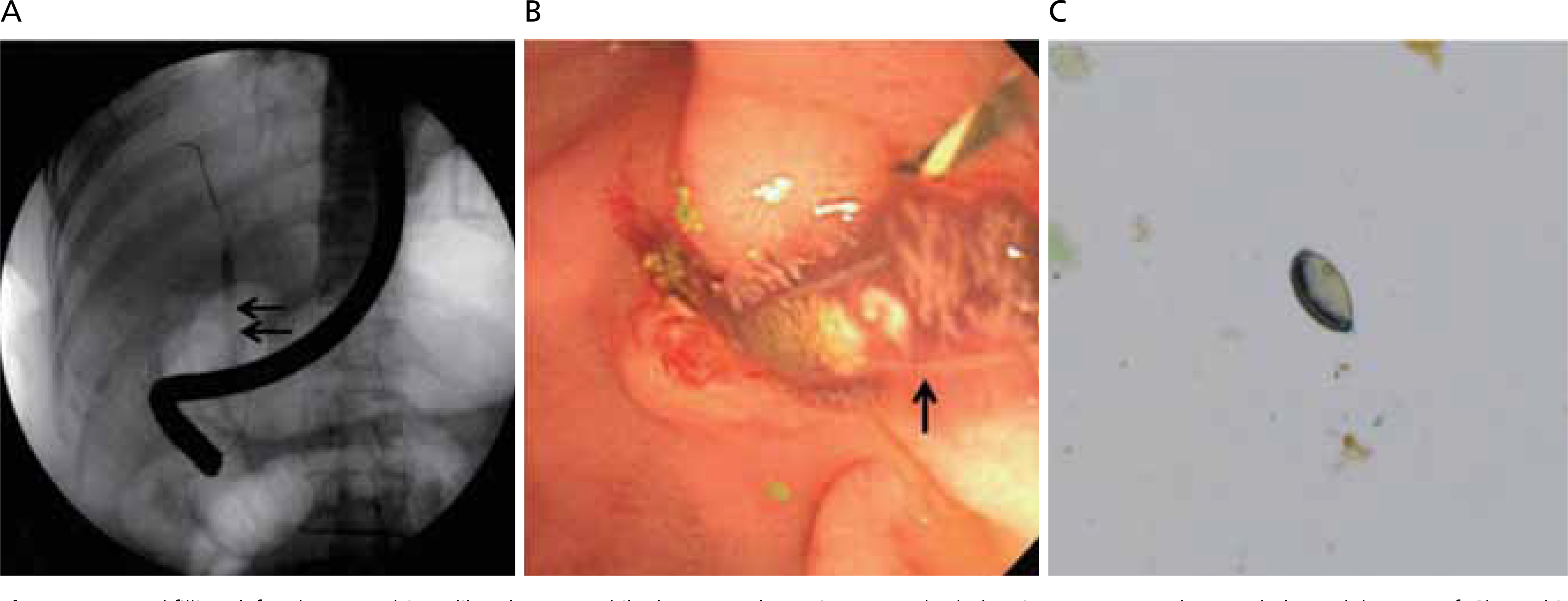
Round filling defect (A, arrows) in a dilated common bile duct on endoscopic retrograde cholangiopancreatography revealed an adult worm of Clonorchis sinensis (B, arrow). Stool examination demonstrated the eggs of C. sinensis seen as yellow-brown ovoid operculation (C, ×400). Reproduced from Yang YM et al. Korean J Pancreas Biliary Tract 2019;24:79-83, according to the Creative Commons license [35].

Enterobius vemicularis eggs obtained by cellphans-tape perianal swab from student. Long diameter of eggs is 60 μm (Photo by Sun Huh).
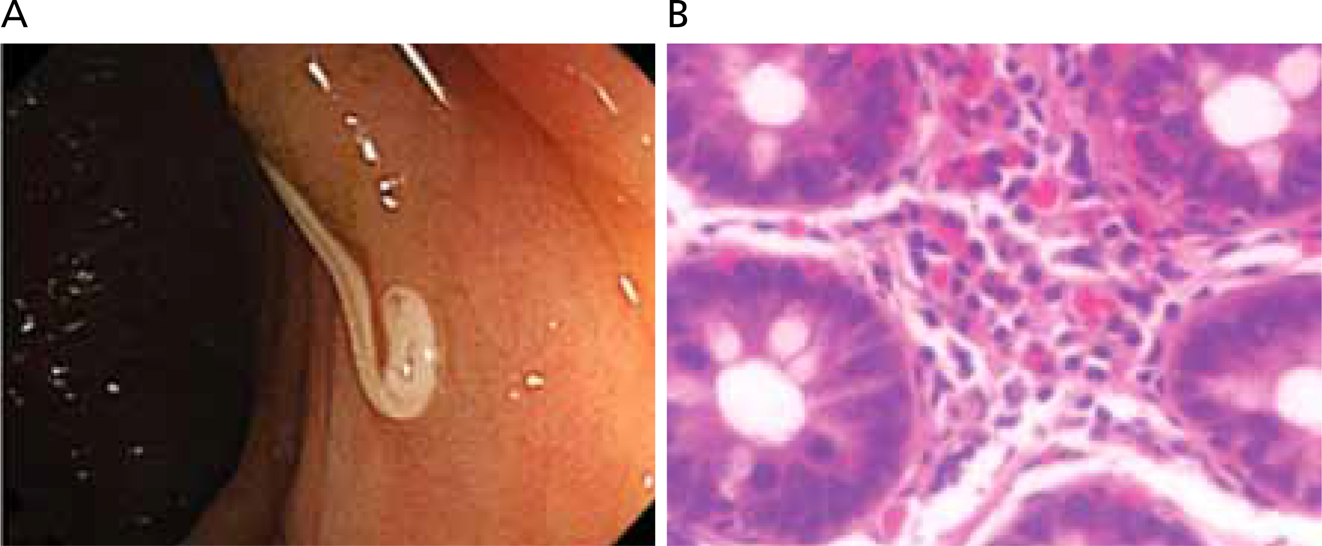
(A) Colonoscopic finding of case 1 showing a whitish worm, Trichuris trichiura, with coiled posterior end embedded in the wall of the cecum. (B) Histologic finding of the cecal mucosa of case 1 which reveals eosinophilic infiltration in the lamina propria (hematoxylin-eosin stain, ×400). Reproduced from Ok KS et al. Korean J Parasitol 2009;47:275-280, according to the Creative Commons license [41].
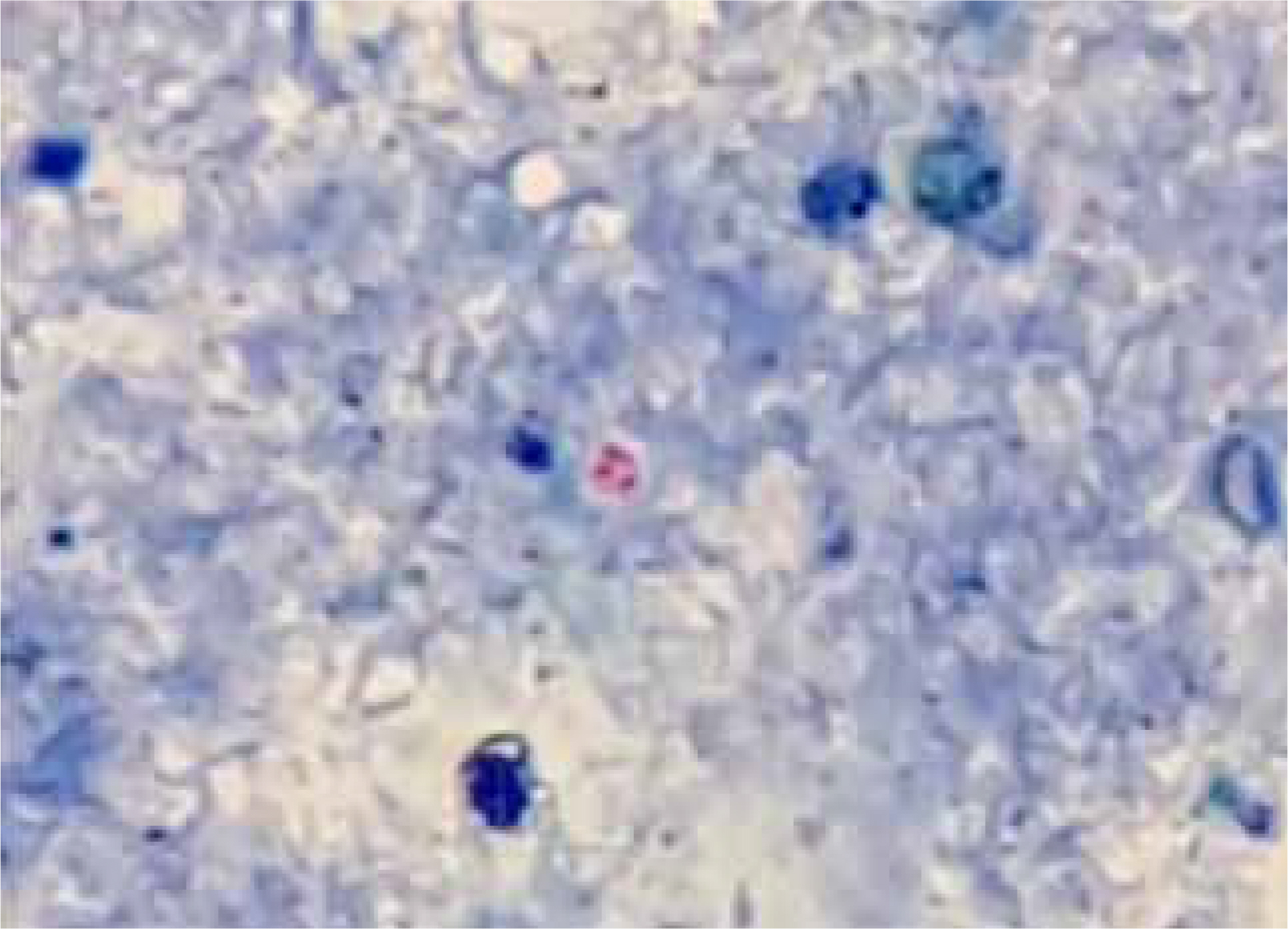
Oocyst of Cryptosporidium parvum obtained from mouse stool expeirmentallly infected with C. parvum. Modified acid-fast stain (×1,000). Four trophozoites are shown as red color in the oocyst of which diameter is 5 μm (Photo by Sun Huh).
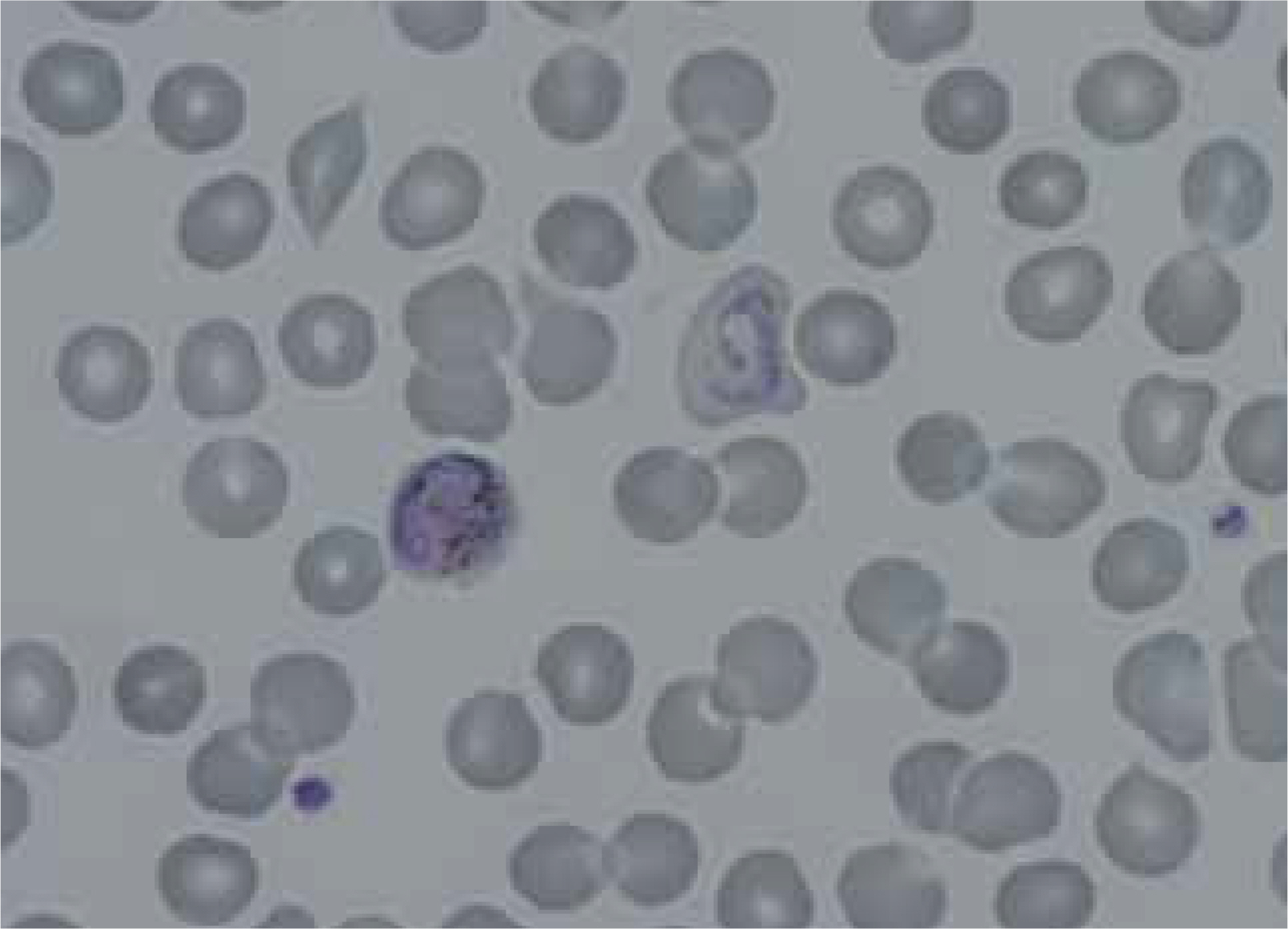
Peripheral blood smear showing a trophozoite and a macrogametocyte of Plasmodium vivax of the patient (Wright-Giemsa stain, ×1,000). Reproduced from Kim NH et al. Korean J Parasitol 2015;53:215-218, according to the Creative Commons license [54].

Classification of malaria cases from 2011 to 2018 in Korea according to origin of the infection and the target population in aboriginal cases. Data from Korea Centers for Disease Control and Prevention. Infectious disease portal [Internet]. Cheongju: Korea Centers for Disease Control and Prevention [3].

Results of 8th national survey of intestinal parasitic infection in 2012, the Republic of Korea

Parasitic diseases reported through Korea Infectious Diseases Surveillance System from 2011 to 2019

Imported parasitic diseases reported through Korea Infectious Diseases Surveillance System from 2010 to 2019