한국 메르스 감염의 역학현황과 공중보건학적 대응 조치 방향
Current epidemiological situation of Middle East respiratory syndrome coronavirus clusters and implications for public health response in South Korea
Article information

Abstract
Since May 20, 2015, when the first case of Middle East respiratory syndrome (MERS) in South Korea was confirmed, the cluster case in South Korea has grown to become the largest observed case following Saudi Arabia within the span of one month. Akin to what was observed in the Middle East, confirmed cases were infected through nosocomial transmission where the cluster is largely limited to patients, healthcare workers, and visitors to patients in healthcare facilities with confirmed cases. A major difference from the outbreaks in the Arabian Peninsula has been the large number of tertiary transmission cases in South Korea, which had reached forty cases by June 12. This observation may suggest that despite the lack of genetic mutation of Middle East respiratory syndrome coronavirus (MERS-CoV) in South Korea, the virus may be behaving differently from that of the Middle East. The higher infectiousness of 'super-spreaders' in South Korea also suggests that this assertion should be under further investigation. Suggestions of inadequate triage in emergency rooms, particularly at Samsung Medical Center which accounts for the most nosocomial infection with 60 cases, have been made by several organizations as the basis for this rapid spread. This, however, does not account for the fact that triage was impossible to implement, since the presence of MERS-CoV in South Korea was unknown during the index patient's stay at the healthcare facilities. This paper aims to identify the key factors in the amplified spread of MERS-CoV in South Korea. The first is the initial failure to confirm diagnosis promptly and to isolate the index case after confirmation of MERS in hospital and the lack of detail in tracking potential exposures in the community of the index case before isolation. The second is the early inadequate measures the Korea Centers for Disease Control and Prevention took in categorizing close contacts. Due to inconsistencies in defining what constitutes close contact, a number of cases were neglected from quarantine and were not subjected to investigation. Finally, confirmed or potential MERS patients were admitted for treatment and observation at medical facilities without adequate disease control measures or rooms, such as ventilated single rooms or airborne precaution rooms. Due to the rigid position that MERS-CoV cannot be transmitted via airborne means, infection control measures has so far neglected evidence that smaller droplets (aerosol) containing the virus can act similar to airborne agents, which may account for the widespread and rapid transmission in a emergency room and a patient's room in hospital. Although the South Korean government expects newly confirmed cases to abate in the coming few weeks, without stringent implementation of clearly defined guidelines to control further transmissions, the cessation of the current trend may continue for an extended period. Additionally, due to the high infection rate of super-spreaders in South Korea, efforts to screen for potential super-spreaders and a thorough investigation of those confirmed to be super-spreaders should be done to quickly identify source of infection, to potentially lower the number of secondary, tertiary transmissions and prevent possible quaternary transmissions.
서론
2015년 5월 20일, 한국에서 중동호흡기증후군(Middle East respiratory syndrome, MERS; 메르스) 첫 확진자가 발생한 직후부터 급속한 증가세로 번지고 있다. 6월 12일 현재, 메르스 확진자 126명, 사망자 11명, 격리자 3,680명으로 증가하였다[1] (Figure 1). 사우디아라비아(확진자 1,028명, 사망 451명)에 이어 세계 2위의 메르스 발병국이 되었다[2]. 226명의 메르스 확진자 중 남성이 74명, 여성이 52명이며, 남성은 40대에서, 여성은 50대에서 많은 확진자가 발생하였다. 10대에서도 1명의 남성 환자가 보고되었다(Figure 2).
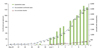
Number of confirmed patients, deaths and quarantines with Middle East respiratory syndrome coronavirus in South Korea (20 May to 12 June, 2015).
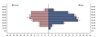
Number of confirmed patients with Middle East respiratory syndrome coronavirus for age and gender in South Korea (20 May to 12 June, 2015).
2015년 6월 12일 현재, 확진자로 보고된 126명의 환자들 중 최초환자로부터 감염된 것으로 판단되는 2차 감염자는 30명으로 전체의 24%인 반면, 3차 감염자는 94명(74%) 이다. 또한 환자와 동일한 병동에 입원하였던 확진자가 62명(49%), 환자의 가족 혹은 보호자 22명(17%) 외에도 환자가 입원한 병원에 방문하였거나 체류하였던 방문자 중에서 감염 확진자도 31명(25%)에 이르고 있다(Figure 3).
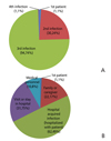
Classification of transmission with route of Middle East respiratory syndrome coronavirus among of all confirmed patients in South Korea (20 May to 12 June, 2015). (A) Classification followed the transmission steps of 126 confirmed patients. (B) Classification followed the characteristics of 126 confirmed patients.
메르스 감염자가 발생한 병원은 서울 강남에 위치한 삼성 서울병원이 60명으로 가장 많고, 평택성모병원이 36명으로 그 뒤를 잇고 있다. 슈퍼전파자로 추정되는 1번, 14번, 16번 환자 중 1번 환자는 확진 전인 5월 15일과 17일 각각 평택 성모병원과 삼성서울병원에 입원하였다. 평택성모병원에서는 5월 20일 이후 36명의 환자가 확진되었다. 또한 14번 환자는 27일에 삼성서울병원에 내원하였으며 삼성서울병원은 6월 4일, 1명의 환자가 발견된 이후 급격한 환자의 증가를 나타내어 현재까지 60명의 환자가 보고되었다. 16번 환자는 5월 25일에 대전에 위치한 대청병원, 그리고 28일에 마찬가지로 대전에 위치한 건양대학교병원에 입원하였으며, 이 두 병원에서 6월 1일부터 6월 12일까지 총 17명의 환자가 감염된 것으로 보고하고 있다(Figure 4).
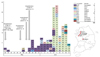
Date of confirmation for Middle East respiratory syndrome coronavirus and distribution of hospitals and locations where patients infected (number of each box means patient number) in South Korea (20 May to 12 June, 2015). MHP, St. Mary's Hospital in Pyeongtaek; YCS, 365 Yeollin Clinic in Seoul; SCA, Seoul Clinic in Asan; KUH, Konyang University Hospital in Daejeon; DCH, Daecheong Hospital in Daejeon; SMC, Samsung Medical Center in Seoul; MHY, St. Mary's Hospital Yeouido in Seoul; AMC, Asan Medical Center in Seoul; HMC, Hallym University Medical Center in Hwaseoung; GMP, Goodmorning Hospital in Pyeongtaek; BHP, Bagae Hospital in Pyeongtaek. Red bold represents the suspected super spreader. a)Death; b)Complete recovery.
한국 내 메르스 확진자들은 의료기관 내 방문 및 체류환자들을 중심으로 한 감염 특성을 보이고 있다. 이는 2014년 사우디아라비아 Jeddah의 메르스 환자들의 주요감염 경로라고 알려진 슈퍼전파자와 병원 내 감염과 유사한 감염경로의 특성을 보이고 있다[3]. 이러한 특성으로 판단컨대, 최초 감염자와 추정 슈퍼전파자의 병원 내 격리가 가장 중요하며 향후 '감염성 질환 관리 및 대책'에 있어서 차단과 격리의 중요한 기준이라고 할 수 있다.
그러나, 한국 보건당국과 일부 전문가들은 MERS-CoV의 공기전파 가능성을 부정하고 직접접촉에 의한 전파 가능성만을 인정하고 있다. 이에 본 저자들은 MERS-CoV의 비말전파와 공기전파에 대한체 과학적 검토와 한국 내 역학현황을 분석하여 향후 차단과 격리범위에 대한 공중보건학적 대응조치를 제시하고자 한다.
에어로졸, 비말의 과학적 정의에 대한 논란
에어로졸(aerosol)은 미세한 고체 또는 액체 방울이 기체에 떠다니는 것을 의미하며 대체로 크기는 0.001 µm에서 100 µm 이다[4]. 과학적인 정의에 따르면 장시간 동안 먼 거리를 부유할 수 있는 작은 크기의 에어로졸은 공기운반입자 (airborne)으로 분류하고 그에 비해 크기가 큰 에어로졸은 비말(droplet)로 분류한다[5].
즉, 에어로졸에 의한 전파는 직접접촉에 의한 비말전파와 공기전파의 두 가지 형태로 일어날 수 있다는 것이다. 비말 전파는 일반적으로 재채기, 기침, 대화 할 때 또는 숨을 내쉴 때 이루어지는데, 이것을 1차 에어로졸화라고 한다[6]. 그에 반해 공기전파는 비말의 수분이 증발하면서 남기는 <5 µm의 비말핵이 퍼지면서 전파되는 것인데, 비말핵은 가벼우며 공기 중에 장시간 부유할 수 있기 때문에 특히 위험할 수 있다[7]. 비말은 표면에 정착한 후에도 진동 또는 기류 등에 의해 바이러스가 다시 공기 중으로 들어가는 2차 에어로졸화 또는 재부유(resuspension)를 일으킬 수 있다[68].
비말과 비말핵은 구형입자의 직경에 따라 공기 중에 떠돌아다니는 시간이 달라지는데 Tellier [9]에 의하면 비말 발생 장소로부터 3 m 이동 범위 내 침강하는데 100 µm의 입자는 4분 20초, 10 µm는 17분, 5 µm는 62분 그리고 3 µm 미만은 거의 침강하지 않고 공기 중에 부유한다고 한다. 또한, 공기 중의 비말은 생성 중에 전하를 가지게 되어 서로 같은 극성의 성질을 띠게 되는데, 이 때문에 각 입자 간에 척력이 발생하여 입자의 공기 중 확산이 일어날 수 있다고 보고한 바 있다[10]. 이와 관련하여 환기시스템과 공기의 흐름은 중증급성호흡기증후군(severe acute respiratory syndrome, SARS), 인플루엔자 같은 전염병의 공기전파와 깊은 연관이 있다[11].
입자의 호흡기 출입경로, 증발 정도에 따른 비말의 크기 변화, 기류에 의한 입자의 이동 경로 등에 따라 감염이 결정되기 때문에 크기에 따른 공기운반입자와 비말의 분류에 관해서는 지속적인 연구가 필요하다[512]. 호흡기 내에서 분열되거나 증식되는 대다수 병원체는 특정 환경이 충족되면 공기전파와 비슷한 양상을 보일 수 있다는 연구결과가 보고되었다[13]. 이와 관련된 실험 연구로 Chao 등[14]은 비말이 평균적으로 기침을 할 때는 11.7 m/s, 말을 할 때는 3.9 m/s 의 빠른 속도로 퍼지는 것을 보고한 바 있다. 비말의 평균 크기는 기침을 할 때 13.5 µm, 말을 할 때 16 µm이며, 관찰 된 비말 중 5 µm 미만의 작은 비말도 존재한다. 이러한 작은 크기의 비말의 경우 빠르게 증발되어 더 작은 크기의 비말핵(droplet nuclei)을 남겨 공기의 흐름을 따라 장시간 부유하며 널리 퍼지는 것으로 보고하였다. 더 나아가 비말핵은 입자가 작기 때문에 오랫동안 공기에 떠다닐 뿐만 아니라 하부 호흡기에 깊숙이 침투 될 수 있다.
앞서 기술한 연구결과를 요약하자면, 5 µm 크기 미만 비말 또는 비말핵은 공기전파의 가능성이 있다는 것이다. 그러나, 2014년 12월 질병관리본부가 발간한 "중동호흡기증후군(MERS) 관리지침(2판)"은 밀접접촉자를 "확진 또는 의심 환자와 신체적 접촉을 한 자(또는 환자가 증상이 있는 동안 2 m 이내의 공간에 1시간 이상 함께 머문자)"로 정의하여, 에어로졸에 의한 공기 중 전파의 원내감염 가능성을 고려하지 않았다. 결과적으로 본 지침에 따른 밀접접촉자의 격리에 관한 한국의 보건복지부와 질병관리본부의 대응조치가 불충분하였음을 시사한다[15]. 미국 질병통제예방센터(Centers for Disease Control and Prevention, CDC)는 밀접접촉(close contact)의 기준을 "1) 6 ft (2 m) 이내 접촉 또는 가운, 장갑, 호흡기, 고글 등의 개인보호장비(personal protective equipment)를 착용하지 않은 상태에서 장기간 동안 입원실 또는 같은 치료 공간 안에 머무른 의료진이나 가족의 경우; 2) 가운, 장갑, 호흡기, 고글 등의 보호장비를 착용하지 않은 상태에서 기침과 같은 전염성 분비물과 직접 접촉한 사람을 의미 한다"고 정의한다[16].
한국질병관리본부의 중동호흡기증후군 (MERS) 대응지침 2판(2014년 12월)은 2015년 5월 24일까지 사용하였으며, 미국 CDC의 밀접접촉 기준 중 개인보호장비를 착용하지 않은 상태에서 장기간 동안 입원실 또는 같은 치료 공간 안에 머무른 의료진이나 가족의 경우를 제외하였다. 결과적으로 이러한 기준은 입원실 또는 같은 치료 공간 내에 머무른 의료진과 가족 중 2 m 이내 공간에 머무른 사람만 접촉 관리 대상에 포함하고 그보다 훨씬 많은 입원실과 응급실의 접촉 관리대상자를 놓치는 결과를 초래한 원인이 되었다. 또한, 관리지침서(2판)의 여러 곳에서 밀접접촉자의 정의가 전반적으로 일정하지 않고 접촉 거리와 시간 등의 세부 기준이 불분명한 것을 지적할 수 있다.
Azhar 등[17]에 따르면 2013년 10월 19일 메르스에 감염된 낙타와 접촉한 사람에게 10월 26일 증상이 발생하였다. 이후 10월 31일 중증의 증상으로 발전하였으며, 11월 6일 같은 환자에게서 MERS-CoV가 검출되었다. 또한, MERS-CoV에 감염된 낙타를 사육하던 낙타농장에서 공기샘플을 사흘간(11월 7일, 8일, 9일) 채취하여 분석한 결과, 11월 7일 MERS-CoV에 감염된 남성과 공기샘플의 MERS-CoV의 유전자 단편이 서로 일치하는 것을 확인한 바 있다.
6월 5일자 Nature News[18]는 병원 내 호흡이 어려운 환자들에게 MERS-CoV 전파가 이루어진 것이 이번 한국에서 메르스 감염의 큰 이유라고 보도한 바 있다. 병원 내 자가호흡이 곤란한 환자들이 호흡보조(aid breathing)를 받을 때 폐에서 에어로졸이 발생할 수 있으며 그 에어로졸에 바이러스가 포함되어 전파될 수 있다. MERS-CoV는 일반적으로 폐 하부를 침범하여 감염되기 때문에 보통 사람들은 기침을 해도 MERS-CoV가 밖으로 나오지 않기 때문에 폐렴환자 등 호흡보조가 필요한 중증의 환자에서는 언제든 에어로졸 형태로 MERS-CoV가 전파될 수 있다는 것을 의미한다. 세계보건기구(World Health Organization, WHO)는 메르스 환자 치료 시 aerosol-generating medical procedures (AGMPs)가 에어로졸을 발생할 수 있다고 보고하고 이에 대한 '공기 전파주의(airborne precaution)'를 권고하고 있다[19].
요약하자면, 병원과 같은 제한된 공간 내에서 메르스 감염자들에게 AGMPs(인공호흡기와 기관 내 삽관, 가래 제거) 등을 수행하는 경우와 MER-CoV의 공기전파 가능성을 인정하고 입원실 또는 같은 치료 공간 안에 머무른 모든 사람들을 밀접접촉 대상자로 관리하는 것이 시급하다.
한국 슈퍼전파자의 역학적 특성
감염전파자의 개인적 특성을 고려한 팔레토 법칙의 양상이 많은 감염성 질환 유행에서 발견 되었다[20]. 이는 전체 감염자 중 20%가 유행 원인의 80%를 차지한다는 가설이며 개인별 전파력이 동일하지 않다는 의미를 가지고 있다. 이와 같이 다른 감염전파자에 비해 상대적으로 많은 숫자의 2차 감염자를 발생시키는 감염자를 슈퍼전파자(super spreader)로 정의하며[21], 현대 감염병 유행의 중요한 특성이다.
슈퍼전파자는 다양한 감염성 질환 유행에서 발견되었다. 2003년 중국 베이징에서 발생한 SARS의 경우 총 77명의 환자 중 66명은 2차 감염을 일으키지 않았으나 3명의 환자가 각각 10명이 넘는 2차 감염자를 발생시켰다[22]. 싱가폴에서는 2003년 SARS에 감염된 전체 238명 중 206명의 병원 감염자 가운데 5명의 슈퍼전파자가 발생하여 한 명이 최대 37명까지 전파시킨 사례가 있었다[232425]. 반면, 베트남의 경우 격리입원기간 동안 전체 33명의 SARS 감염자 중 슈퍼 전파자가 관찰되지 않았으며, 추가적인 2차 감염자 또한 발생하지 않았다[26].
슈퍼전파는 병원이라는 특수한 환경에서 일어날 가능성이 크다는 보고가 있다. 2014년 Jeddah에서 MERS-CoV 양성으로 나온 케이스와 병원 기록을 모두 분석한 연구에서는 증상을 보인 191명 메르스 양성 환자 중 20.9%가 의료진이었으며 의료진이 아닌 나머지 151명의 97.3%가 증상 발현 14일 전 병원에 다녀간 경험이 있다. 따라서 2014년 Jeddah 메르스 2차 감염은 병원에서 현저하게 증가했으며 MERS-CoV에 쉽게 감염될 수 있는 환자를 감별하는 것이 중요하다고 보고하였다.
슈퍼전파를 발생시키는 원인은 아직까지 명확하지 않으나, 최근 연구에서 동시감염이 중요한 관리요인임을 알려졌다. 1960년대 미국에서 유행한 신생아 포도상구균 감염사례에서 슈퍼전파자들은 공통적으로 상기도 감염증상을 보였고[27], 1983년 같은 지역의 포도상구균성 피부감염 사례에서도 동일한 결과가 나타났다[28]. 또한 2005년 미국 웨이크 포레스트 대학에서 지원자 11명을 대상으로 한 실험연구는 rhinovirus 감염이 포도상구균 보균자의 감염력을 최소 2배에서 최대 34배까지 증가시키는 결과를 보여주었다[29]. 동시감염이 감염력을 증가시키는 메커니즘이 명확하게 밝혀지지는 않았으나 Sherertz 등[30]은 포도상 구균의 체내 균락 (colony) 존재와 상기도 감염이 비강의 협소와 유체역학적 특성을 초래하고 결과적으로 비말의 에어로졸화 증가와 환자의 비말 분출을 증가시켰다고 주장하였다.
그밖에도 숙주, 병인 및 환경요인에 따라 슈퍼전파자의 발생 위험을 추정해볼 수 있다. Table 1은 현재까지 발생한 슈퍼전파 발생 사례를 토대로 관련요인을 기술한 자료이다 [31]. 메르스는 WHO [32]가 Cauchemez 등 [33]의 연구결과를 인용하여 보고한 기초감염재생산수(R0)가 0.8-1.3이었으나, 이미 우리나라에서 63명 이상을 감염시킨 슈퍼전파 추정 사례가 발생하고 있다. 이들의 발생 양상을 분석하여 슈퍼전파 가능 의심 사례를 빠른 시일 내 찾아 조치하는 것이야말로 이번 메르스 감염 전파관리의 핵심이라고 볼 수 있다. Li 등[34]은 감염병 증상이 발현된 시점으로부터 입원이 4일 이상 지연될 경우 슈퍼전파의 발생 가능성이 높아진다고 주장하였다.
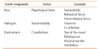
Super-spreading events are shaped by host, pathogen, and environmental factors
이번 국내 메르스 유행에서도 슈퍼전파의 양상이 나타나고 있다. 따라서 슈퍼전파자의 발생 양상을 분석하여 이들을 적시에 찾아 조치하는 것이야말로 이번 메르스 유행 관리의 핵심 사안이라고 볼 수 있다. 한국 내 최초 MERS-CoV 감염자가 내원하였던 서울의 한 병원과 아산의 한 병원에서 2명의 의료진을 통한 2차 감염이 일어났으며, 평택성모병원에서 27건의 2차 감염을 초래하였다. 1번 환자로부터 감염된 것으로 판단되는 15번 환자는 경기도 화성의 한림대학교병원으로 이동, 의료인을 포함한 4명의 3차 감염자를 발생시킨 것으로 보인다. 또한 메르스 확산에 있어 주요한 슈퍼전파자로 추정되는 14번 환자의 경우 평택성모병원에서 확진 판정을 받은 후 삼성서울병원으로 이동한 환자이며, 6월 12일 현재까지 삼성서울병원에서 발생한 60명과 평택 굿모닝병원 3명의 3차 감염 원인을 제공한 것으로 여겨진다. 마찬가지로 평택 성모병원의 16번 환자는 대전에 소재한 두 병원(건양대학교 병원, 대청병원)에서 17명의 환자에 대한 전파를 야기하였다(Figure 5). 이러한 역학적 현황을 볼 때, 향후 추가적인 슈퍼 전파자의 감염과 당분간 3차 감염자의 추가 보고가 이어질 가능성이 있으며 대량의 4차 감염자 발생 가능성에 대한 우려를 주목할 필요가 있다.
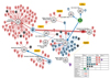
Distribution of transmission of Middle East respiratory syndrome coronavirus clusters and suspected super spreader in South Korea (20 May to 12 June, 2015).
한국과 중동에서 발병한 메르스 환자의 특성
Table 2는 한국에서 발생하는 메르스의 발병양상과 사우디아라비아의 발병양상의 차이를 정리한 내용이다[353637]. Breban 등[35]은 여러 명의 환자가 2차 감염을 시키지 않고 한 환자가 다수에게 2차 감염을 시킨다는 가정 하에서 prepandemic SARS 기초감염재생산수(basic reproduction number, R0)는 0.8인 반면 MERS-CoV의 R0는 0.69 (95% CI, 0.50-0.92)라고 보고하였으며, 한 환자가 소수에게 2차 감염을 시키고 그러한 환자가 여러 명이 있다는 가정 하에서는 MERS-CoV의 R0는 0.60 (0.42-0.80)로 보고한 바 있다
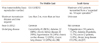
Differences between Middle East respiratory syndrome outbreak in the Middle East and South Korea
최초 메르스 환자가 병원에서 발생했을 때 한국 보건복지부는 기존 기초감염재생산수 0.6-0.8 자료에 근거하여 우리나라에서 감염력이 높지 않을 것이라고 발표하였으며[1538], 이로 인하여 슈퍼관리자에 대한 관리 필요성을 간과한 것으로 보인다. 본 연구자들은 Breban 등[35]의 첫 번째 연구가설을 적용하여 기초감염재생산수를 잠정적으로 산출하였다. 6월 11일을 기준으로 126명의 확진자중 감염경로가 파악이 되지 않은 1명의 환자를 제외한 후 2 세대까지의 기초감염재생산수를 산출한 결과 약 4.0명으로 추정되었다. 이는 현재까지 보고된 극히 제한적인 역학자료를 바탕으로 계산된 추정치이며 향후 정확한 자료를 바탕으로 추가적인 연구가 필요하다. 한국에서 확진자가 증가하면서 메르스 감염환자들의 일반적인 증상인 고열을 보이지 않는 경우에서 MERS-CoV 양성 확진을 진단받는 경우가 발생하였다. 또한, 고위험군 집단이 취약집단으로 알려져 있었으나, 한국의 메르스 감염자 중 36.2%는 기저질환이 없는 건강한 사람들이었다. Assiri 등[36]은 사우디아라비아에서 발생한 47명의 MERS-CoV 감염환자에 대해 역학적·임상적 특성을 분석하여 메르스 확진환자의 기저 질환을 동반상병 45명(96%), 당뇨병 32명(68%), 만성 신장질환 23명(49%), 고혈압 16명(34%) 만성 심질환 13명(28%), 만성 폐질환 12명(26%) 등의 순으로 보고한 바 있다. 한국 보건복지부의 발표자료에 따르면 한국 내 메르스 확진자 45명을 분석한 결과, 기저질환이 없는 건강한 사람은 21명(36.2%)이라고 발표하였다. 또한, 메르스 확진자의 기저질환은 고혈압 10명(17.2%), 당뇨병 8명(13.7%), 암 7명(12.0%), 만성 폐질환 6명(10.3%) 등의 순으로 보고하였다[37].
결론 및 공중보건학적 대응 시사점
Azhar 등[17]이 보고한 메르스 감염환자가 소유한 낙타 농장에서 환자의 동일한 MERS-CoV 입자가 발견되었으나 MERS-CoV 입자가 감염력을 갖고 있는지에 대해서는 아직 명확한 증거는 없다. 메르스의 공기매개 여부와 공기 중에 떠 있는 상황에서 MERS-CoV의 감염력이 얼마나 지속 되는지 등에 대한 과학적인 근거는 아직 충분하지 않은 상황이다. 그럼에도 불구하고 본 저자들이 '병원 내 공기전파 가능성을 배제해서는 안된다'고 주장하는 이유는 다음과 같다. 감염성질환의 원인과 경로 등이 과학적으로 엄밀하게 입증된 증거가 없는 경우라 하더라도 예견 가능한 연구 보고들을 토대로 'precautionary principle'에 의거하여 공중보건학적 예방 관리 대책를 수립하고 시행하는 것이 가장 중요한 기본 원리이기 때문이다.
Harriman 등[39]과 미국 CDC [40]는 '입원환자에 대한 감염예방관리 권고(Interim infection prevention and control recommendations for hospitalized patients with Middle East respiratory syndrome coronavirus (MERS-CoV)'를 통해 병원 전파경로 등에 관한 정보가 과학적으로 확실하지 않은 상황에서는 precautionary principle에 따라 모든 메르스 감염환자들에 대해 airborne precautions(예: 수술용 마스크가 아닌 N-95 마스크, 보호의 및 고글 등)을 권고하고 있다.
SARS는 발병 당시 비말감염으로 간주하였으나 비행기 안에서 감염자 좌석 기준 7줄 앞에 자리했던 승객이 감염되거나, 같은 호텔의 같은 층을 사용했던 손님 중 환자가 다수 발생한 사례, 홍콩의 한 아파트 단지에서 1,000명 이상의 발병 사례가 보고되는 등, 각종 관련 사례들이 보고되어 공기감염으로 감염될 수 있다고 판단하였다[6].
그러나, 상기 SARS의 경우는 지역사회에서의 공기감염을 위험성을 강조하고자 한 것이다. SARS와 메르스는 모두 병원 내 공기감염의 위험성 인정과 그에 따른 airborne precaution의 적용은 일치하나, 메르스의 경우 SARS와는 달리 지역사회에서의 공기감염 위험성에 대한 명백한 역학적 증거가 없는 것은 사실이다. 따라서 메르스가 지역사회 내에서 는 공기감염의 우려가 없다고 하는 것은 적절하다. 그러나 병원 내 제한된 공간 내에서의 비말과 에어로졸의 공기전파에 의한 감염 가능성을 입증할 수 있는 다양한 역학적 그리고 실험적 연구결과들을 고려할 때, MERS-CoV의 병원 내 공기감염의 가능성을 부정하는 것은 부적절하다.
2015년 6월 12일 현재까지 한국 내 발병양상과 역학자료를 분석한 결과, 한국 내 메르스 감염의 초기 대응에 있어서 문제점, 최초 환자의 격리와 실패 그리고 소위 슈퍼감염자로 부터의 2차 감염의 증가에 대한 본 저자들의 견해는 다음과 같다.
첫째, 국내 메르스 첫 환자 발생 직후, 2014년 12월 작성 된 매뉴얼 '중동호흡기증후군(MERS) 관리지침(2판)'[15]에 따라 대응을 하였으나 결과적으로 선제적인 격리차단 대응이 실패한 것으로 보인다. '메르스 대응지침(2판)'에 따르면 밀접접촉자의 기준을 "의심 환자와 신체적 접촉을 한 자(또는 환자가 증상이 있는 동안 2 m 이내의 공간에 1시간 이상 함께 머문자)"로 한정함으로 인하여, 작은 크기의 비말 혹은 에어로졸의 동일 공간 내(의료기관 내) 공기전파로 인한 2차 감염자의 발생을 차단하지 못한 원인이 되었다. 실제 이는 14번 환자로부터 삼성서울병원 응급실 내 의료인력과 방문환자가 감염되는 계기가 되었다.
둘째, 밀접접촉자들에 대한 격리차단의 실패이다.
미국 CDC [41]는 메르스 감염자 혹은 접촉자 중 병원입원이 필요하지 않은 사람들에 대한 자가격리 실행지침(Implementing home care and isolation or quarantine of people not requiring hospitalization for MERS-CoV)을 통해 의료전문가는 자가격리 이전에 주 또는 지방보건부와 의 관리자들과 점검 후 주거환경이 자가격리에 적당한지 그리고 CDC interim guidance를 따를 수 있는 환경인지 평가하여야 한다고 기술하고 있다. 그러나, 한국은 자가격리를 실시하기 전에 밀접접촉자들의 주거환경요인 등을 사전에 검토하거나 격리자에 대한 교육과 지원이 이루어지지 않았다. 2015년 6월 3일자 Nature News는 한국에서 MERSCoV에 처음 감염된 68세 남성은 5월 11일부터 확진 받은 5월 20일 전까지의 시간동안 다른 네 곳의 병원을 방문하였으며 그로 인해 병원 내 MERS-CoV 확산을 초래한 것으로 보고하였으며[18], The Lancet News(2015년 6월 13일)[42]는 밀접접촉자 중의 한 사람이 자가격리 지시를 거부하고 중국을 방문한 사실을 언급하면서 밀접접촉자에 대한 격리 관리가 적절하지 않았음을 지적한 바 있다.
셋째, 무엇보다도 한국에서의 가장 중요하고 치명적인 문제점은 슈퍼전파자에 대한 추가적인 감염 차단과 격리 실패 이었다. 결과적으로 최초 환자(1번 환자, index case)에 대한 병원 내 격리 실패로 27명의 2차 감염 발생, 14번 환자는 응급실 내 체류 3일간 격리 조치가 이루어지지 않아 63명 추가 감염 발생 그리고 16번 환자는 17명에게 메르스 2차 감염을 전파하는 상황을 초래하였다.
넷째, 2차 병원 내 감염성질환 관리체계의 미흡한 역량과 미흡한 환기시설 등의 환경적 요인이다. 유럽 질병예방통제센터(European Centre for Disease Prevention and Control)[2]는 한국 내 메르스 감염환자의 확산원인으로 호흡기 증상을 보이는 환자가 응급실 내원 시 여행력 확보 등의 미흡한 환자 분류 체계임을 보고하였다. 또한, The Lancet News (2015년 6월 13일)[42]는 한국에서의 메르스 발생은 중동 이외의 지역에서 일어난 가장 큰 규모의 집단발병 사례로 이는 사우디아라비아의 사례와 마찬가지로 불충분한 병원감염관리 역량이 한국에서의 전파의 주요 원인으로 보고한 바 있다.
다섯째, 에어로졸과 미세 비말로 인한 공기매개 전파가능성을 배제하고 비말에 의한 직접 접촉 감염 경우만을 고려한 것은 초동대응 단계에서 최초 환자의 격리가 실패를 하게 된 가장 중요한 원인이었다고 추정된다. 그러나 현재까지 한국 정부는 메르스 확진자가 집단 발생한 병원 내에서 공기 전파의 가능성을 부인하고 비말의 직접접촉으로 인한 감염만을 공식적으로 인정하고 있다. 이와 같은 감염방식에 대한 정부의 지나친 경직적인 기준으로 인하여 직접 접촉에 의한 감염 의심자 이외의 추가적 감염 의심자가 관리되지 않을 가능성이 여전히 존재한다.
마지막으로, 정부의 미흡한 위기관리 소통이라고 할 수 있다. 대응초기 의료기관에게 메르스 확진환자의 정보와, 경유병원과 확진병원 등에 대한 정보공개가 지연되어 또 다른 2차, 3차 감염이 발생하였다. 실제, 삼성서울병원의 경우 응급실 내 음압격리 병실과 선별진료소가 있음에도 불구하고, MERS-CoV 감염환자의 접촉여부에 대한 정보 공유가 신속하고 투명하게 되지 않아 응급실내에서의 격리가 시행 되지 않았다. 따라서 한국-WHO 메르스 합동평가단이 권고한 바와 같이, 효과적인 위기관리소통체계가 시급히 필요하다[43].
한국 내 메르스 감염자의 확산에 대한 본 연구자들의 견해는 현재까지 알려진 제한된 역학 자료를 토대로 도출한 잠정적인 연구결과이다. 향후 한국에서 MERS-CoV 감염에 관한 추가적인 역학 연구 정보들과 관련 연구 결과들이 도출된 다면 본 연구자들의 견해는 지지되거나 수정될 수 있다. 그럼에도 불구하고 본 연구자들의 견해는 '감염성 질환 예방 및 관리에 있어 사전예방 원칙'의 중요성을 강조하고 현 시점에서 시급한 공중보건학적 대응관리의 방향을 제언하는데 의의가 있다.
Acknowledgement
We would like to thank the researchers at the Institute for Occupational and Environmental Health at Korea University (Seung-Hun Ryu, Eunsun Lee, Jiwook Park, Minjoo Ku, Min Soo Kim, Byeo Ri Lee, Jiwon Moon, Minhee Kim, and Tinyami Eric Tandi) for their academic support.