Low Back Pain: Review of Anatomy and Pathophysiology

Article information

J Korean Med Assoc. 2006;49(8):656-664
Publication date (electronic) : 2006 August 31
doi : https://doi.org/10.5124/jkma.2006.49.8.656
Department of Anesthesiology and Pain Medicine, Pusan University College of Medicine, Korea. shinsw@pusan.ac.kr

Abstract

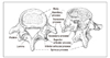
Most of the structures in the lumbar region including the visceral organs could be the sources of low back pain. The management of low back pain starts from a thorough understanding of the anatomical structures and the underlying pathophysiologic processes related to the generation of the pain. Mechanical stresses applying to the lumbar spine and the inflammatory changes contribute to the generation of low back pain. Many nerves branching from the spinal and autonomic nerves supply all of the musculoskeletal structures in the lumbar area. There are extensive nociceptive nerve fibers in the facet joints and some small fibers in the outer layer of discs and ligaments of the lumbar vertebrae. They respond to the mechanical, chemical and other stimuli. Acute pain caused by tissue trauma or inflammation is well controlled by the removal or elimination of its causes. In idiopathic, uncontrolled and chronic pain, however, the long-lasting nociceptive stimuli and many chemical mediators released from the tissue injury and inflammation sensitize the local nervous system. They change the normal process of pain transmission to neuropathic pain. For the proper treatment of low back pain, not only the knowledge of anatomical structures but also the understanding of the pathophysiology of chronic neuropathic pain is necessary.

References

1. Finch PM, Taylor JM. In : Waldman SD, ed. Functional anatomy of the spine. Interventional pain management 2001. 2nd edth ed. Philadelphia: WB Saunders; 43–71.
2. Czerniecki JM, Goldstein B. In : Loeser JD, ed. General considerations of pain in the low back, hips, and lower extremities. Bonica's management of pain 2001. 3rd edth ed. Philadelphia: Lippincott Williams & Wilkins; 1475–1507.
3. McCulloch JA, Transfeldt EE. Macnab's backache 1998. 3rd edth ed. Baltimore: Williams & Wilkins; 1–74.
6. Cavanaugh JM, Ozaktay AC, Yamashita T, Avramov A, Getchell TV, King AI. Mechanisms of low back pain. Clin Orthop Relat Res 1997. 335166–180.
7. Manchikanti L. Facet joint pain and the role of neural blockade in its management. Curr Rev Pain 1999. 3(5)348–358.
8. Jang YH, Lee JK, Chen JK, Chung JK. A clinical survey of patients of pain clinic. J Korean Pain Soc 1995. 8103–109.

Article information Continued

Figure 1

Lumbar vertebra

Figure 2

Nerve supply of cross section of lumbar spine

IVD: intervertebral disc, VB: vertebral body, all: anterior longitudinal ligament, pll: posterior longitudinal ligament, st: sympathetic trunk, grc: gray ramus communicans, svn: sinovertebral nerve, vr: ventral ramus, dr: dorsal ramus, ds: dural sac, zj: zygapophyseal joint, m: muscle (Bogduk, 1991)

Figure 3

Segmental nerve supply of lumbar spine (Stanley, 1983)

Table 1

Causes of low back pain

Table 1

Table 2

Pathophysiology of intervertebral disc aging and degeneration

Table 2

Table 3

Common chemical substances in disc degeneration and their functions

Table 3

MMP: matrix metalloproteinase, IL: interleukin, TNF: tumor necrosis factor, PG: prostaglandin, CGRP: calcitonin gene related peptide, sP: substance P, TIMP: tissue inhibitor of metalloproteinase, TGF: transforming growth factor, EGF: insulin-like growth factor, PDGF: platelet-derived growth factor