Diagnosis and non-operative treatment of shoulder pain

Article information

J Korean Med Assoc. 2019;62(12):629-635
Publication date (electronic) : 2019 December 12
doi : https://doi.org/10.5124/jkma.2019.62.12.629
Department of Physical Medicine and Rehabilitation, Seoul Metropolitan Government-Seoul National University Boramae Medical Center, Seoul National University College of Medicine, Seoul, Korea.
Corresponding author: Shi-Uk Lee. shiuk.lee@gmail.com
Received 2019 October 15; Accepted 2019 November 18.

Abstract

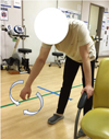
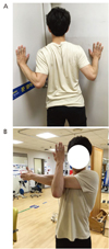
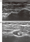
The shoulder is the third most common region in which musculoskeletal pain occurs, involving symptoms that cause a decrease in quality of life and a reduced ability to perform activities of daily life if untreated. Many lesions cause shoulder pain; therefore, we included those commonly encountered in primary care clinics. We aimed to describe the diagnosis and non-operative or conservative treatment modalities of rotator cuff tendinopathy, impingement syndrome, rotator cuff tear, adhesive capsulitis, biceps tendonitis, and acromioclavicular arthritis. Exercise has been reported to be effective for the treatment of impingement syndrome, and the effect of exercise has been shown to reduce pain. Both home-based self-exercise and exercise supervised by professionals are equally effective. Types of exercise should include stretching, active, active-assistive, and resistive exercise. Ten repetitions per set for 3 sets of each exercise should be prescribed initially post-injury. Repetition and resistance should be increased as strength grows. The use of ultrasonography for the diagnosis of shoulder pain has increased recently. Corticosteroid injection under ultrasonographic guidance is effective in reducing pain.

Acknowledgement

Special thanks to Sang Yoon Lee, MD, PhD and Jung Woo Kim, MD (Seoul National University Boramae Medical Center, Depart-ment of Physical Medicine & Rehabilitation) for the help on the photographs in the article.

Notes

Conflict of Interest

No potential conflict of interest relevant to this article was reported.

References

1. Urwin M, Symmons D, Allison T, Brammah T, Busby H, Roxby M, Simmons A, Williams G. Estimating the burden of musculoskeletal disorders in the community: the comparative prevalence of symptoms at different anatomical sites, and the relation to social deprivation. Ann Rheum Dis 1998;57:649–655.
2. Laslett LL, Burnet SP, Jones JA, Redmond CL, McNeil JD. Musculoskeletal morbidity: the growing burden of shoulder pain and disability and poor quality of life in diabetic outpatients. Clin Exp Rheumatol 2007;25:422–429.
3. Neer CS 2nd. Impingement lesions. Clin Orthop Relat Res 1983;(173):70–77.
4. Bigliani LU, Levine WN. Subacromial impingement syndrome. J Bone Joint Surg Am 1997;79:1854–1868.
5. Burbank KM, Stevenson JH, Czarnecki GR, Dorfman J. Chronic shoulder pain: part I. Evaluation and diagnosis. Am Fam Physician 2008;77:453–460.
6. Naredo E, Aguado P, De Miguel E, Uson J, Mayordomo L, Gijon-Baños J, Martin-Mola E. Painful shoulder: comparison of physical examination and ultrasonographic findings. Ann Rheum Dis 2002;61:132–136.
7. Arroll B, Goodyear-Smith F. Corticosteroid injections for painful shoulder: a meta-analysis. Br J Gen Pract 2005;55:224–228.
8. Kuhn JE. Exercise in the treatment of rotator cuff impingement: a systematic review and a synthesized evidence-based rehabilitation protocol. J Shoulder Elbow Surg 2009;18:138–160.
9. Neer CS 2nd. Anterior acromioplasty for the chronic impingement syndrome in the shoulder. 1972. J Bone Joint Surg Am 2005;87:1399.
10. Murrell GA, Walton JR. Diagnosis of rotator cuff tears. Lancet 2001;357:769–770.
11. Burk DL Jr, Karasick D, Kurtz AB, Mitchell DG, Rifkin MD, Miller CL, Levy DW, Fenlin JM, Bartolozzi AR. Rotator cuff tears: prospective comparison of MR imaging with arthrography, sonography, and surgery. AJR Am J Roentgenol 1989;153:87–92.
12. Jacobson JA. Fundamentals of musculoskeletal ultrasound 2nd edth ed. Philadelphia: Elsevier/Saunders; 2013.
13. Redler LH, Dennis ER. Treatment of adhesive capsulitis of the shoulder. J Am Acad Orthop Surg 2019;27:e544–e554.
14. Arkkila PE, Kantola IM, Viikari JS, Rönnemaa T. Shoulder capsulitis in type I and II diabetic patients: association with diabetic complications and related diseases. Ann Rheum Dis 1996;55:907–914.
15. Lee HJ. Differential diagnosis of common shoulder pain. J Korean Med Assoc 2014;57:653–660.
16. Nam HS, Lee SU. Conservative management of shoulder pain with common causes. J Korean Med Assoc 2014;57:661–666.
17. Rizk TE, Gavant ML, Pinals RS. Treatment of adhesive capsulitis (frozen shoulder) with arthrographic capsular distension and rupture. Arch Phys Med Rehabil 1994;75:803–807.

Article information Continued

Figure 1

An ultrasonography image indicating tendinopathy of the supraspi-natus tendon.

Figure 2

Pendulum exercise for the shoulder. Informed consent was obtained from the study participant.

Figure 3

(A) Anterior shoulder stretching exercise. (B) Posterior shoulder stretching exercise. Informed consent was obtained from the subject.

Figure 4

(A) Internal and external rotator strengthening exercise using an elastic band. (B) Scaption exercise of the shoulder. Informed consent was obtained from the participant.

Figure 5

(A) Chair press. (B) Upright rows. Informed consent was obtained from the participant.

Figure 6

Ultrasonography of a supraspinatus tendon tear. (A) Normal tendon. (B) Articular side partial tear. (C) Bursal side partial tear. (D) Full thickness tear.

Figure 7

Intraarticular injection in the shoulder under ultrasonographic guidance.

Figure 8

Ultrasonographic image of the biceps brachii tendon, transverse plane. (A) Normal. (B) Effusion.