


 |
 |
| J Korean Med Assoc > Volume 52(7); 2009 > Article |
Abstract
Multiple sclerosis (MS) is an inflammatory autoimmune disorder of the central nervous system (CNS) and one of the most common disabling neurological diseases of young adults. Although the exact mechanisms involved in MS pathogenesis remain unclear, MS is believed to be caused by interactions between as yet unidentified environmental factors and susceptibility genes. Symptoms commonly occurred in MS include visual disturbance; weakness; spasticity; sensory disturbances; ataxia; bladder, bowel, and sexual dysfunction; fatigue; affective symptoms; and cognitive impairment. Most patients initially undergo a relapsing-remitting course, however, without treatment, the majority of them make a transition to the secondary progressive form. The clinical diagnosis is based on demonstrating neurological lesions, predominantly in the white matter, that are disseminated over space with the lapse of time. The key to the successful MS management is to prevent disability. Although there is no effective cure for MS, therapies are available that mitigate the course of the disease, treat relapses and improve symptoms, all of which place a significant impact on patients' quality of life. Recent clinical trials suggest that early identification and treatment are critical to optimize the treatment benefit. Currently six agents have been specifically approved for mitigating the course of MS. These include three formulations of interferon beta, glatiramer acetate, mitoxantrone, and natalizumab. Recent advances in understanding of immune pathogenesis lead us to new therapeutic approaches focused on precise target mechanisms. Many ongoing clinical trials will provide better treatment protocols in near future.
References
1. Ropper AH, Brown RH. Adams and Victor's principles of neurology 2005;8th ed. New York: McGraw-Hill. 771-796.
2. Compston A. Genetic epidemiology of multiple sclerosis. J Neurol Neurosurg Psychiatry 1997;62:553-561.
3. Kurtzke JF. Mutliple sclerosis in time and space-geographic clues to cause. J Neurovirol 2000;6:134-140.
4. Raghuwanshi A, Joshi SS, Christakos S. Vitamin D and multiple sclerosis. J Cell Biochem 2008;105:338-343.
5. Gale CR, Martyn CN. Migrant studies in multiple sclerosis. Prog Neurobiol 1995;47:425-448.
6. Sibley WA, Bamford CR, Clark K. Clinical viral infections and multiple sclerosis. Lancet 1985;1:1313-1315.
7. Willer CJ, Dyment DA, Risch NJ, Sadovnick AD, Ebers GC. Canadian Collaborative Study Group. Twin concordance and sibling recurrence rates in multiple sclerosis. Proc Natl Acad Sci USA 2000;100:12877-12882.
8. Olerup O, Hillert J. HLA class II-associated genetic susceptibility in multiple sclerosis: a critical evaluation. Tissue Antigens 1991;38:1-15.
9. Svejgaard A. The immunogenetics of multiple sclerosis. Immunogenetics 2008;60:275-286.
10. Chofflon M. Mechanisms of action for treatments in multiple sclerosis; Does a heterogeneous disease demand a multi-targeted therapeutic approach? BioDrugs 2005;19:299-308.
11. Bar-Or A. Immunology of multiple sclerosis. Neurol Clin 2005;23:149-175.
12. Archelos JJ, Storch MK, Hartung HP. The role of B cells and autoantibodies in multiple sclerosis. Ann Neurol 2000;47:694-706.
13. Link H, Muller R. Immunoglobulins in multiple sclerosis and infections of the nervous system. Arch Neurol 1971;25:326-344.
14. Johnson KP, Nelson BJ. Multiple sclerosis: diagnostic usefulness of cerebrospinal fluid. Ann Neurol 1977;2:425-431.
15. Raine CS, Cannella B, Hause SL, Genain CP. Demyelination in primate autoimmune encephalopmyelitis and acute multiple sclerosis lesion: a case for antigen-speficit antibody mediation. Ann Neurol 1999;46:144-160.
16. Lucchinetti C, Bruck W, Parisi J, Scheithauer B, Rodriguez M, Lassmann H. Heterogeneity of multiple sclerosis lesion: implications for the pathogenesis of demyelination. Ann Neurol 2000;47:707-717.
17. Buntinx M, Moreels M, Vandenabeele F, Lambrichts I, Raus J, Steels P, Stinissen P, Amellot M. Cytokine-induced cell death in human oligodendroglial cell lines: I. Synergistic effects of IFN-gamma and TNF-alpha on apoptosis. J Neurosci Res 2004;76:834-845.
18. Trapp BD, Peterson J, Ransohoff RM, Rudick R, Mork S, Bo L. Axonal transection in the lesions of multiple sclerosis. N Engl J Med 1998;29:278-285.
19. Hauser SL, Goodin DS. In: Braunwald E, Fauci AS, Kasper DL, Hauser SL, Longo DL, Jameson JL, Isselbacher K, editor. Multiple sclerosis and other demyelinating diseases. Harrison's Online 2006;New York: McGraw-Hill. Available from: URL:
http://www.accessmedicine.com
20. Rizzo JF III, Lessell S. Risk of developing multiple sclerosis after uncomplicated optic neuritis: A long-term prospective study. Neurology 1988;38:185-190.
21. Optic neuritis study group. The five-year risk of MS after optic neuritis. Experience of the optic neuritis treatment trial. Neurology 1997;49:1404-1413.
22. Lucchinetti CF, Kiers L, O'Duffy A, Gomez MR, Cross S, Leavitt JA, O'Brien P, Rodriguez M. Risk factors for developing multiple sclerosis after childhood optic neuritis. Neurology 1997;49:1413-1418.
23. Beck RW, Trobe JD, Moke PS, Gal RL, Xing D, Bhatti MT, Brodsky MC, Buckley EG, Chrousos GA, Corbett J, Eggenberger E, Goodwin JA, Katz B, Kaufman DI, Keltner JL, Kupersmith MJ, Miller NR, Nazarian S, Orengo-Nania S, Savino PJ, Shults WT, Smith CH, Wall M. Optic neuritis study group. High-and low-risk profiles for the development of multiple sclerosis within 10 years after optic neuritis: experience of the optic neuritis treatment trial. Arch Ophthalmol 2003;121:944-949.
24. Lublin FD, Reingold SC. Defining the clinical course of multiple sclerosis: results of an international survey. National Multiple Sclerosis Society (USA) Advisory Committee on Clinical Trials on New Agents in Multiple Sclerosis. Neurology 1996;46:907-911.
25. Polman CH, Reingold SC, Edan G, Filippi M, Hartung HP, Kappos L, Lublin FD, Metz LM, McFarland HF, O'Connor PW, Wandberg-Wollheim M, Thompson AJ, Weinchenker BG, Wolinsky JS. Diagnostic criteria for multiple sclerosis: 2005 revisions to the "McDonald Criteria". Ann Neurol 2005;58:840-846.
26. Katzman GL. In: Osborn AG, Blaser SI, Salzman KL, Katzman GL, Provenzale J, Castillo M, Hedlund GL, Illner A, Harnsberger HR, Cooper JA, Jones BV, Hamilton BE, editor. Multiple sclerosis. Diagnostic imaging. Brain 2004;Salt Lake City: Amirsys. I-8.74-I-8.77.
27. Jiang H, Milo R, Swoveland P, Johnson KP, Panitch H, Dhib-Jalbut S. Interferon beta-1b reduces interferon gamma-induced antigen-presenting capacity of human glial and B cells. J Neuroimmunol 1995;61:17-25.
28. Genc K, Dona DL, Reder AT. Increased CD80 (+) B cells in active multiple sclerosis and reversal by interferon beta-1b therapy. J Clin Invest 1997;99:2664-2671.
29. Teleshova N, Bao W, Kivisakk P, Ozenci V, Mustafa M, Link H. Elevated CD40 ligand expressing blood T-cell levels in multiple sclerosis are reversed by interferon-beta treatment. Scand J Immunol 2000;51:312-320.
30. Sharief MK, Semra YK, Seidi OA, Zoukos Y. Interferon-beta therapy downregulates the anti-apoptosis protein FLIP in T cells from patients with multiple sclerosis. J Neuroimmunol 2001;120:199-207.
31. Calabresi PA, Pelfrey CM, Tranquill LR, Maloni H, McFarland HF. VLA-4 expression on peripheral blood lymphocytes is downregulated after treatment of multiple sclerosis with interferon beta. Neurology 1997;49:1111-1116.
32. Calabresi PA, Tranquill LR, Dambrosia JM, Stone LA, Maloni H, Bash CN, Frank JA, McFarland HF. Increases in soluble VCAM-1 correlate with a decrease in MRI lesions in multiple sclerosis treated with interferon beta-1b. Ann Neurol 1997;41:669-674.
33. Trojano M, Avolio C, Liuzzi GM, Ruggieri M, Defazio G, Liguori M, Santacroce MP, Paolicelli D, Giuliani F, Riccio P, Livrea P. Changes of serum sICAM-1 and MMP-9 induced by rIFNbeta-1b treatment in relapsing-remitting MS. Neurology 1999;53:1402-1408.
34. Calabresi PA, Stone LA, Bash CN, Frank JA, McFarland HF. Interferon beta results in immediate reduction of contrast-enhanced MRI lesions in multiple sclerosis patients followed by weekly MRI. Neurology 1997;48:1446-1448.
35. Petersen B, Bendtzen K, Koch-Henriksen N, Ravnborg M, Ross C, Sorensen PS. Danish multiple sclerosis group. Persistence of neutralizeing antibodies after discontinuation of IFN beta therapy in patients with relapsing-remitting multiple sclerosis. Mult Scler 2006;12:247-252.
36. Jacobs LD, Beck RW, Simon JH, Kinkel RP, Brownscheidle CM, Murray TJ, Simonian NA, Slasor PJ, Sandrock AW. CHAMPS Study Group. Intramuscular interferon beta-1a therapy initiated during a first demyelinating event in multiple sclerosis. N Engl J Med 2000;343:898-904.
37. Comi G, Filippi M, Barkhof F, Durelli L, Edan G, Fernández O, Hartung H, Seeldrayers P, Sørensen PS, Rovaris M, Martinelli V, Hommes OR. Early Treatment of Multiple Sclerosis Study Group. Effect of early interferon treatment on conversion to definite multiple sclerosis: a randomised study. Lancet 2001;357:1576-1582.
38. Kappos L, Polman CH, Freedman MS, Edan G, Hartung HP, Miller DH, Montalban X, Barkhof F, Bauer L, Jakobs P, Pohl C, Sandbrink R. Treatment with interferon beta-1b delays conversion to clinically definite and McDonald MS in patients with clinically isolated syndromes. Neurology 2006;67:1242-1249.
39. Arnon R. The development of Cop 1 (Copaxone), and innovative drug for the treatment of multiple sclerosis: personal reflextions. Immunol Lett 1996;50:1-15.
40. Teitelbaum D, Meshorer A, Hirshfeld T, Arnon R, Sela M. Suppression of experimental allergic encephalomyelitis by a synthetic polypeptide. Eur J Immunol 1971;1:242-248.
41. Neuhaus O, Farina C, Wekerle H, Hohlfeld R. Mechanisms of action of glatiramer acetate in multiple sclerosis. Neurology 2001;56:702-708.
42. Weber MS, Starck M, Wagenpfeil S, Meinl E, Hohlfeld R, Farina C. Multiple sclerosis: glatiramer acetate inhibits monocyte reactivity in vitro and in vivo. Brain 2004;127:1370-1378.
43. Kim HJ, Ifergan I, Antel JP, Seguin R, Duddy M, Lapierre Y, Jalili F, Bar-Or A. Type 2 monocyte and microglia differentiation mediated by glatiramer acetate therapy in patients with multiple sclerosis. J Immunol 2004;172:7144-7153.
44. Fox EJ. Mechanism of action of mitoxantrone. Neurology 2004;63:15-18.
45. Niino M, Bodner C, Simard ML, Alatab S, Gano D, Kim HJ, Trigueiro M, Racicot D, Guérette C, Antel JP, Fournier A, Grand'Maison F, Bar-Or A. Natalizumab effects on immune cell responses in multiple sclerosis. Ann Neurol 2006;59:748-754.
46. Miller DH, Khan OA, Sheremata WA, Blumhardt LD, Rice GP, Libonati MA, Willmer-Hulme AJ, Dalton CM, Miszkiel KA, O'Connor PW. International natalizumab multiple sclerosis trial group. A controlled trial of natalizumab for relapsing multiple sclerosis. N Engl J Med 2003;348:15-23.
47. Kleinschmidt-DeMasters BK, Tyler KL. Progressive multifocal leukoencephalopathy complicating treatment with natalizumab and interferon-b-1a for multiple sclerosis. N Engl J Med 2005;353:369-374.
48. Langer-Gould A, Atlas SW, Green AJ, Bollen AW, Pelletier D. Progressive multifocal leukoencephalopathy after natalizumab therapy for Crohn's disease. N Engl J Med 2005;353:362-368.
49. Buttmann M, Rieckmann P. Treating multiple sclerosis with monoclonal antibodies. Expert Rev Neurother 2008;8:433-455.
50. Cohen BA, Reickmann P. Emerging oral therapies for multiple sclerosis. Int J Clin Pract 2007;61:1922-1930.
51. Kesselring J, Beer S. Symptomatic therapy and neurorehabilitation in multiple sclerosis. Lancet Neurol 2005;4:643-652.
Figure 1
APC = antigen presenting cell, IFN = interferon, MBP = myelin basic protein, MHC = major histocompatibility complex, MMP = matrix metalloproteinase, NOI = nitric oxide intermediates, ROI = reactive oxygen intermediates, TCR = T cell receptor, TNF = tumor necrosis factor, VCAM = vascular cell adhesion molecule, VLA = very late antigen.
The five key immunopathogenic processes in multiple sclerosis (MS) targeted by MS therapies (10). (A) T cell activation and differentiation into T-helper (Th) -1 cells, (B) interleukin (IL)-2-induced proliferation of activated Th1 cells, (C) recruitment of B cells and monocytes by activated Th1 cells, (D) activated Th1 cell trafficking across the blood- brain barrier (BBB), (E) T cell reactivation and induction of immune cell-mediated demyelination.
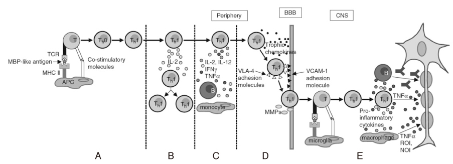
Figure 3
Representing MRI of multiple sclerosis.
(A) Fluid-attenuated inversion recovery (FLAIR) MRI demonstrates typical multiple perpendicular-axis periventricular white matter lesions called "Dawson's fingers".
(B) Gadolinium enhanced T1-weighted MRI shows well-enhancing MS plaque.

Table 1
2005 Revisions to the McDonald Diagnostic Criteria for Multiple Sclerosis (25)
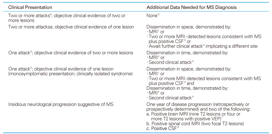
If criteria indicated are fulfilled and there is no better explanation for the clinical presentation, the diagnosis is MS; if suspicious, but the criteria are not completely met, the diagnosis is "possible MS" ; if another diagnosis arises during the evaluation that better explains the entire clinical presentation, then the diagnosis is "not MS".
a An attack is defined as an episode of neurological disturbance for which causative lesions are likely to be inflammatory and demyelinating in nature. There should be subjective report (backed up by objective findings) or objective observation that the event lasts for at least 24 hours.
b No additional tests are required; however, if tests (MRI, CSF) are undertaken and are negative, extreme caution needs to be taken before making a diagnosis of MS. Alternative diagnoses must be considered. There must be no better explanation for the clinical picture and some objective evidence to support a diagnosis of MS.
c MRI demonstration of space dissemination must fulfill the criteria derived from Barkhof and colleagues and Tintoré and coworkers.
d Positive CSF determined by oligoclonal bands detected by established methods (isoelectric focusing) different from any such bands in serum, or by an increased IgG index.
e MRI demonstration of time dissemination must fulfill the criteria in Table 3.
f Abnormal VEP of the type seen in MS.
MS = multiple sclerosis, MRI = magnetic resonance imaging, CSF = cerebrospinal fluid, VEP = visual- evoked potential
Table 2
MRI criteria to demonstrate brain abnormality and demonstration of dissemination in space
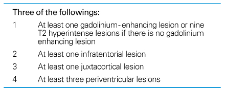
NOTE: A spinal cord lesion can be considered equivalent to a brain infratentorial lesion: an enhancing spinal cord lesion is considered to be equivalent to an enhancing brain lesion, and individual spinal cord lesions can contribute together with individual brain lesions to reach the required number of T2 lesions.
Table 4
List of US food and drug administration-approved disease-modifying therapies
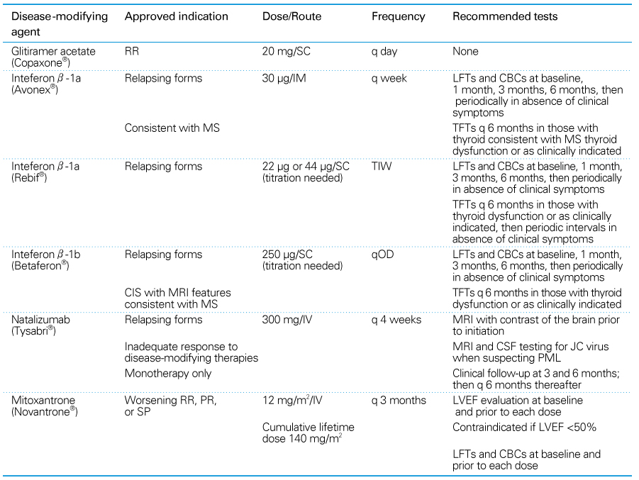
CBC = complete blood count with platelets, CIS = clinically isolated syndrome, DMT = disease-modifying therapy, LFT = liver function tests, LVEF = left ventricular ejection fraction, MS = multiple sclerosis, PML = progressive multifocal leukoencephalopathy, PR = progressive-relapsing, q = every, qOD = every other day, RR = relapsing-remitting, SC = subcutaneous, SP = secondary progressive, TFT = thyroid function tests, TIW = 3 times a week
- TOOLS
-
- Share :
-


-
METRICS

-
Related articles in
J Korean Med Assoc -
Multidrug-resistant Tuberculosis1998 May;41(5)
Multiple Scrotal Nodules1999 April;42(4)
Multidrug-resistant Tuberculosis2006 September;49(9)
- Editorial Office
-
37 Ichon-ro 46-gil, Yongsan-gu, Seoul
Tel: +82-2-6350-6562 Fax: +82-2-792-5208 E-mail: jkmamaster@gmail.com
Copyright © 2024 by Korean Medical Association.
