


 |
 |
| J Korean Med Assoc > Volume 52(2); 2009 > Article |
Abstract
Angiogenesis, the process whereby new capillaries are formed by outgrowth from existing microvessels, is required for tumor growth and metastasis, as well as for healing of ischemic injuries. Because angiogenesis is a promising target for molecular therapies, there is a real need to develop molecular imaging methods to monitor angiogenesis activity. Direct imaging of angiogenesis can help define the pathophysiology of angiogenic processes in vivo, and foster personized medicine by identifying patients likely to respond to angiogenesis-targeted drugs and accurately monitor the therapeutic efficacy. Promising imaging targets include integrins, vascular endothelial growth factor (VEGF) receptors, and matrix metalloproteinases. While MRI and optical imaging modalities are also workable, radiolabeled RGD (arginine-glycine-aspartate) probes that target αvβ3 integrins overexpressed on activated endothelia are the most extensively investigated and successful angiogenesis imaging technique to date. This technique has repeatedly been validated in preclinical models of cancers and ischemic diseases, and clinical studies are presently ongoing to elucidate the value of RGD positron image tomography (PET) imaging in human patients. Herein, we review the current status of angiogenesis imaging research with special emphasis on integrin-targeted techniques.
References
1. Ferrara N, Kerbel RS. Angiogenesis as a therapeutic target. Nature 2005;438:967-974.
2. Kerbel R, Folkman J. Clinical translation of angiogenesis inhibitors. Nat Rev Cancer 2002;2:727-739.
3. Carmeliet P, Jain RK. Angiogenesis in cancer and other diseases. Nature 2000;407:249-257.
4. Carmeliet P. Angiogenesis in health and disease. Nat Med 2003;9:653-660.
5. Tongers J, Roncalli JG, Losordo DW. Therapeutic angiogenesis for critical limb ischemia: microvascular therapies coming of age. Circulation 2008;118:9-16.
6. Ahn A, Frishman WH, Gutwein A, Passeri J, Nelson M. Therapeutic angiogenesis: a new treatment approach for ischemic heart disease-part I. Cardiol Rev 2008;16:163-171.
7. Provenzale JM. Imaging of angiogenesis: clinical techniques and novel imaging methods. Am J Roentgenol 2007;188:11-23.
8. McDonald DM, Choyke PL. Imaging of angiogenesis: from microscope to clinic. Nat Med 2003;9:713-725.
9. Riggs TL. Research and development costs for drugs. Lancet 2004;363:184.
10. Cai W, Rao J, Gambhir SS, Chen X. How molecular imaging is speeding up antiangiogenic drug development. Mol Cancer Ther 2006;5:2624-2633.
11. Bergers G, Benjamin LE. Tumorigenesis and the angiogenic switch. Nat Rev Cancer 2003;3:401-410.
12. Ferrara N, Gerber HP, LeCouter J. The biology of VEGF and its receptors. Nat Med 2003;9:669-676.
13. Hwang R, Varner J. The role of integrins in tumor angiogenesis. Hematol Oncol Clin North Am 2004;18:991-1006.
14. Sipkins DA, Cheresh DA, Kazemi MR, Nevin LM, Bednarski MD, Li KCP. Detection of tumor angiogenesis in vivo by αvβ3 targeted magnetic resonance imaging. Nat Med 1998;4:623-626.
15. Brooks PC, Clark RA, Cheresh DA. Requirement of vascular integrin αvβ3 for angiogenesis. Science 1994;264:569-571.
16. Haubner R, Wester HJ, Reuning U, Senekowitsch-Schmidtke R, Diefenbach B, Kessler H, Stöcklin G, Schwaiger M. Radiolabeled αvβ3 integrin antagonists: a new class of tracers for tumor targeting. J Nucl Med 1999;40:1061-1071.
17. Haubner R, Wester HJ, Burkhart F, Senekowitsch-Schmidtke R, Weber W, Goodman SL, Kessler H, Schwaiger M. Glycosylated RGD-containing peptides: tracer for tumor targeting and angiogenesis imaging with improved biokinetics. J Nucl Med 2001;42:326-336.
18. Haubner R, Wester HJ, Weber WA, Mang C, Ziegler SI, Goodman SL, Senekowitsch-Schmidtke R, Kessler H, Schwaiger M. Noninvasive imaging of αvβ3 integrin expression using 18F-labeled RGD-containing glyco-peptide and positron emission tomography. Cancer Res 2001;61:1781-1785.
19. Pichler BJ, Kneilling M, Haubner R, Braumüller H, Schwaiger M, Röcken M, Weber WA. Imaging of delayed-type hypersensitivity reaction by PET and 18F-galacto-RGD. J Nucl Med 2005;46:184-189.
20. Haubner R, Weber WA, Beer AJ, Vabuliene E, Reim D, Sarbia M, Becker KF, Goebel M, Hein R, Wester HJ, Kessler H, Schwaiger M. Noninvasive visualization of the activated αvβ3 integrin in cancer patients by positron emission tomography and [18F]galactoRGD. PLoS Med 2005;2:e70.
21. Beer AJ, Haubner R, Goebel M, Luderschmidt S, Spilker ME, Wester HJ, Weber WA, Schwaiger M. Biodistribution and pharmacokinetics of the αvβ3 selective tracer 18F-galacto-RGD in cancer patients. J Nucl Med 2005;46:1333-1341.
22. Beer AJ, Grosu AL, Carlsen J, Kolk A, Sarbia M, Stangier I, Watzlowik P, Wester HJ, Haubner R, Schwaiger M. [18F]galacto-RGD positron emission tomography for imaging of αvβ3 expression on the neovasculature in patients with squamous cell carcinoma of the head and neck. Clin Cancer Res 2007;13:6610-6616.
23. Mulder WJ, van der Schaft DW, Hautvast PA, Strijkers GJ, Koning GA, Storm G, Mayo KH, Griffioen AW, Nicolay K. Early in vivo assessment of angiostatic therapy efficacy by molecular MRI. FASEB J 2007;21:378-383.
24. Zhang C, Jugold M, Woenne EC, Lammers T, Mor-genstern B, Mueller MM, Zentgraf H, Bock M, Eisenhut M, Semmler W, Kiessling F. Specific targeting of tumor angiogenesis by RGD-conjugated ultrasmall super-paramagnetic iron oxide particles using a clinical 1.5-T magnetic resonance scanner. Cancer Res 2007;67:1555-1562.
25. Lee BC, Sung HJ, Kim JS, Jung KH, Choe YS, Lee KH, Chi DY. Synthesis of Tc-99m labeled glucosamino-Asp-cyclic (Arg-Gly-Asp-d-Phe-Lys) as a potential angiogenesis imaging agent. Bioorg Med Chem 2007;15:7755-7764.
26. Jung KH, Lee KH, Paik JY, Ko BH, Bae JS, Lee BC, Sung HJ, Kim DH, Choe YS, Chi DY. Favorable biokinetic and tumor-targeting properties of 99mTc-labeled glucosamino RGD and effect of paclitaxel therapy. J Nucl Med 2006;47:2000-2007.
27. Lee KH, Jung KH, Song SH, Kim DH, Lee BC, Sung HJ, Han YM, Choe YS, Chi DY, Kim BT. Radiolabeled RGD uptake and αv integrin expression is enhanced in ischemic murine hindlimbs. J Nucl Med 2005;46:472-478.
28. Hua J, Dobrucki LW, Daseghi MM, Zhang J, Bourke BN, Cavaliere P, Song J, Chow C, Jahanshad N, van Royden N, Buschmann I, Madri JA, Mendisabal M, Sinusas AJ. Noninvasive imaging of angiogenesis woth a 99mTc labeled peptide targeted at αvβ3 integrin after murine hindlimb ischemia. Circulation 2005;111:3255-3260.
29. Meoli D, Sadeghi M, Krassilnikova S, Bourke BN, Giordano FJ, Dione DP, Su H, Edwards DS, Liu S, Harris TD, Madri JA, Zaret BL, Sinusas AJ. Noninvasive imaging of myocardial angiogenesis following experimental myocardial infarction. J Clin Invest 2004;113:1684-1691.
30. Makowski MR, Ebersberger U, Nekolla S, Schwaiger M. In vivo molecular imaging of angiogenesis, targeting alphavbeta3 integrin expression, in a patient after acute myocardial infarction. Eur Heart J 2008;29:2201.
31. Li S, Peck-Radosavljevic M, Kienast O, Preitfellner J, Hamilton G, Kurtaran A, Pirich C, Angelberger P, Dudczak R. Imaging gastrointestinal tumours using vascular endothelial growth factor-165 (VEGF165) receptor scintigraphy. Ann Oncol 2003;14:1274-1277.
32. Backer MV, Levashova Z, Patel V, Jehning BT, Claffey K, Blankenberg FG, Backer JM. Molecular imaging of VEGF receptors in angiogenic vasculature with single-chain VEGF-based probes. Nat Med 2007;13:504-509.
33. Lu E, Wagner WR, Schellenberger U, Abraham JA, Klibanov AL, Woulfe SR, Csikari MM, Fischer D, Schreiner GF, Brandenburger GH, Villanueva FS. Targeted in vivo labeling of receptors for vascular endothelial growth factor: approach to identification of ischemic tissue. Circulation 2003;108:97-103.
34. Li S, Peck-Radosavljevic M, Kienast O, Preitfellner J, Havlik E, Schima W, Traub-Weidinger T, Graf S, Beheshti M, Schmid M, Angelberger P, Dudczak R. Iodine-123-vascular endothelial growth factor-165 (123I-VEGF165). Biodistribution, safety and radiation dosimetry in patients with pancreatic carcinoma. Q J Nucl Med Mol Imaging 2004;48:198-206.
Figure 1
Integrin-targeted angiogenesis imaging strategy.
(A) Heterodimeric αvβ3 integrin receptors overexpressed on the surface of activated endothelial cells. The extracellular domain of the protein recognizes RGD tripeptide sequences as the binding motif.
(B) General structure of integrin-targeting imaging probes that contain cyclic-RGD residues.
(C, D) Scintigraphic images after injection of a 99mTc labeled cyclic-RGD probe in (C) a murine tumor model showing clear uptake of a carcinoma and (D) a rat myocardial infarction model showing focal uptake in the cardiac region of ischemia.
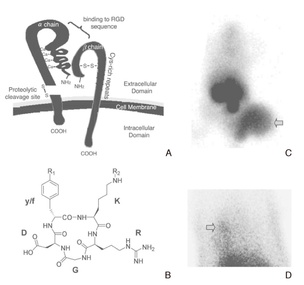
Figure 2
Clinical RGD PET images in human patients.
(A) RGD PET/MRI fusion image of a patient with squamous cell carcinoma in the right oral cavity showing intense lesion uptake. The left hand shows transaxial images and the right hand shows corresponding 3D reconstruction images. Adapted by permission from the American Association for Cancer Research: Clin Cancer Res (22), copyright (2007).
(B) MRI, perfusion PET/CT, and RGD PET/CT images of a patient with acute infarction of the apex and apico-anterior myocardial wall. Focal increased uptake of 18F labeled cyclic-RGD is shown in the area of infarction (right) where perfusion (middle) and systolic function (left) is reduced. Adapted by permission from Oxford University Press: Eur Heart J (30), copyright (2008).
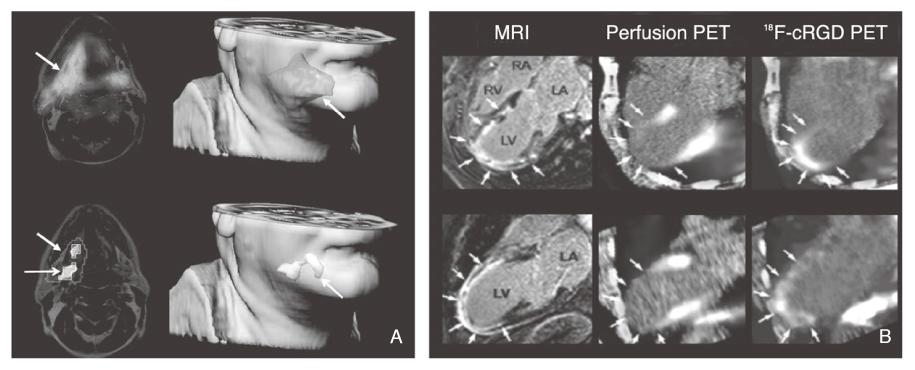
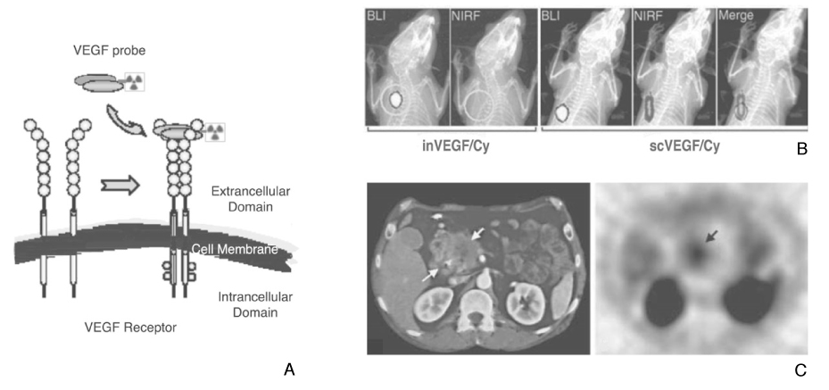
Figure 3
VEGF receptor-targeted angiogenesis imaging.
(A) VEGF probes bind to the extracellular domain of VEGF receptors overexpressed on activated endothelial cells.
(B) Optical images after injection a Cy.5.5 labeled VEGF probe in a mouse with a luciferase-expressing tumor transplant shows tumor delineation by both luminescent and fluorescent signals. Adapted by permission from Macmilla Publishers Ltd: Nat Med (32), copyright (2007).
(C) Transaxial CT and 123I-VEGF images in a patient with primary pancreatic adenocarcimoma showing clear uptake of the malignant lesion. Adapted by permission from Edizioni Minerva Medica: Q J Nucl Med Mol Imaging (34), copyright (2004).
- TOOLS
-
- Share :
-


-
METRICS

-
- 0 Crossref
- Scopus
- 1,129 View
- 5 Download
-

-
Related articles in
J Korean Med Assoc -
Molecular Genetic Aspects of Alzheimer's Disease2002 April;45(4)
Molecular MR Imaging2004 February;47(2)
Gene Therapy and Molecular Imaging2004 February;47(2)
Molecular Imaging of Atherosclerosis2009 February;52(2)
Molecular Imaging in Neurodegenerative Diseases2009 February;52(2)
- Editorial Office
-
37 Ichon-ro 46-gil, Yongsan-gu, Seoul
Tel: +82-2-6350-6562 Fax: +82-2-792-5208 E-mail: jkmamaster@gmail.com
Copyright © 2024 by Korean Medical Association.
