


 |
 |
| J Korean Med Assoc > Volume 49(12); 2006 > Article |
Abstract
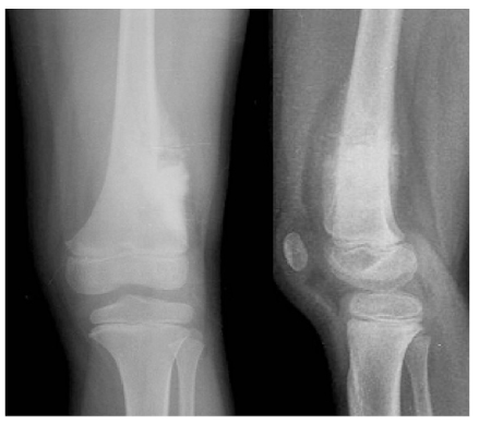
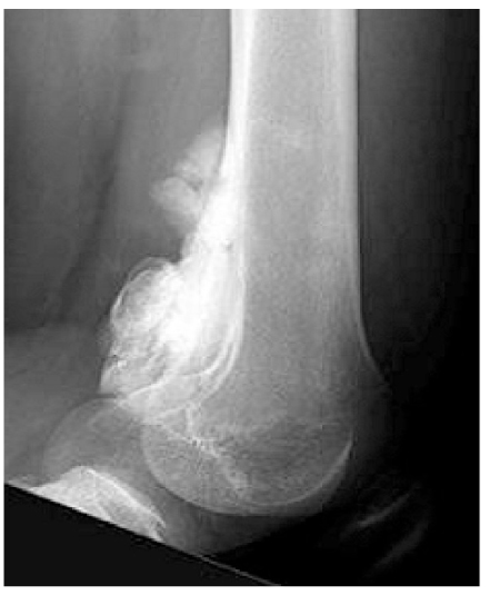
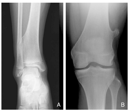
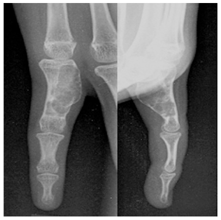
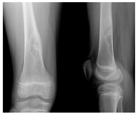
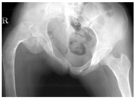
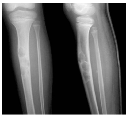
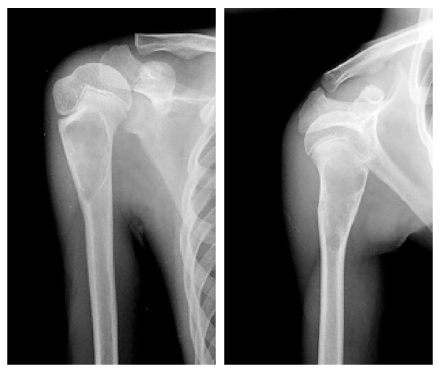
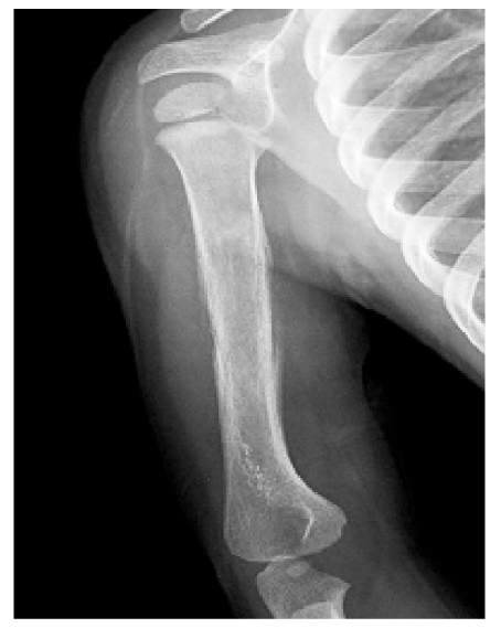
Primary bone tumors in pediatric age group are uncommon, and even when they do occur, they are usually benign. The primary malignant tumors that occur predominantly in children are two bone tumors, namely, osteosarcoma and Ewing's sarcoma. An adequate history and physical examination are the first and most important steps in evaluating a patient with a bone tumor. All suspected bone tumors should be evaluated initially with plain roentgenograms. Then the additional diagnostic studies, such as computed tomography(CT), magnetic resonance imaging(MRI) and technetium bone scan can be used, if necessary. Biopsy should be the last step in evaluation. Most of benign bone tumors usually do not require treatment other than a periodic follow-up evaluation. The optimal treatment of the malignant bone tumor often requires a combination of radiation therapy, chemotherapy, and wide surgical excision or amputation. Early detection of a malignant bone tumor not only may make the difference between life and death but also may allow successful salvage surgery rather than amputation of the limb.
References
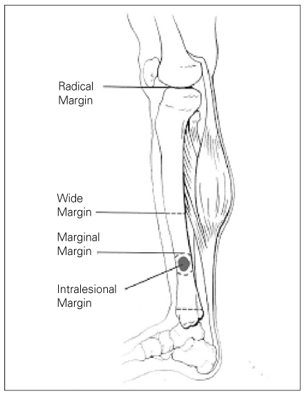
1. Enneking W. Musculoskeletal tumor surgery 1983;New York: Churchill Livingstone.
2. Madwell JE, Ragsdale BD, Sweet DE. Radiologic and pathologic analysis of solitary bone lesions: Part I-Internal margins. Radiol Clin North Am 1981;19:715-748.
3. Ragsdale BD, Madewell JE, Sweet DE. Radilogic and pathologic analysis of solitary bone lesions: Part II-periosteal reactions. Radiol Clin North Am 1981;19:749-783.
4. Springfield DS, Gebghardt MC. In: Morrissy RT, Weinstein SL, editor. Bone and soft tissue tumors. Lovell and Winter's Pediatric Orthopaedics 2001;5th edition. Philadelphia: Lippincott Williams & Willinkins. 507-562.
5. In: Heinrich SD, Scarborough MT, editor. Pediatric Orthopaedic Oncology. Orthop Clin North Am 1996.
6. Jaramillo D, Laor T, Gebhardt MC. Pediatric musculoskeletal neoplasms: evaluation with MR imaging. Magn Reson Imaging Clin N Am 1996;4:749-770.
7. Mankin HJ, Lange TA, Spanier S. The hazards of the biopsy in patients with malignant primary bone and soft-tissue tumors. J Bone Joint Surg Am 1982;64:1121-1127.
8. Mankin Hj, Mankin CJ, Simon MA. The hazards of the biopsy, revisited. Members of the Musculoskeletal Tumor Society. J Bone Joint Surg Am 1996;78:656-663.
9. Simon MA, Biermann JS. Biopsy of bone and soft-tissue lesions. J Bone Joint Surg Am 1993;75:616-621.
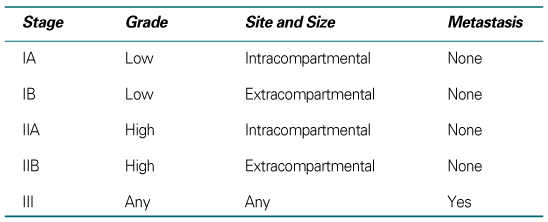
10. Enneking WF, Spanier SS, Goodman MA. A system for the surgical staging of musculoskeletal sarcoma. Clin Orthop Relat Res 1980;153:106-120.
11. Huvos AG. Bon tumors: Diagnosis, Treatment, and Prognosis 1991;2nd ed. Philadelphia: WB Saunders.
12. Greenspan A, Remagen W. Differential Diagnosis of Tumors and Tumor like Lesions of Bone and Joints 1998;Philadelphia: Lippincott-Raven.
13. Himelstein BP, Dormans JP. Malignant bone tumors of childhood. Pediatr Clin North Am 1996;43:967-984.
14. Rougraff BT, Simon MA, Kneisl JS, Greenberg DB, Mankin HJ. Limb salvage compared with amputation for osteosarcoma of the distal end of the femur: a long-term ontological, functional, and quality-of-life study. J Bone Joint Surg Am 1994;76:649-656.
15. Copley L, Dormans JP. Benign pediatric bone tumors: Evaluation and treatment. Pediatr Clin North Am 1996;43:949-966.
16. Dormans JP, Flynn JM. In: Beaty JH, Kasser JR, editor. Pathologic fracture associated with tumors and unique conditions of the muscoloskeletal edition. Rockwood and Wilkin's Fractures in Children 2001;5th ed. Philadelphia: Lippincott Williams & Wilkins.
17. Dormans JP, Pill SG. Fractures through bone cysts: Unicameral bone cysts, aneurysmal bone cysts, fibrous cortical defects, and nonossifying fibromas. Instr Course Lect 2002;51:457-467.
18. Grabias SL, Campbell CJ. Fibrous dysplasia. Orthop Clin North Am 1977;8:771-783.
19. Martinez V, Sissons HA. Aneurysmal bone cyst: a review of 123 cases including primary lesions and those secondary to other bone pathology. Cancer 1988;61:2291-2304.
20. Szendroi M, Arato G, Ezzati A, Huttl K, Szavcsur P. Aneurysmal bone cyst: its pathogenesis based on angiographic, immunohistochemical and eletron microscopic studies. Pathol Oncol Res 1998;4:277-281.
21. Dormans JP, Hanna BG, Johnston DR, Khurana JS. Surgical treatment and recurrence rate of aneurysmal bone cysts in children. Clin Orthop 2004;421:205-211.
22. Sessa S, Sommelet D, Lascombes P, Prevot J. Treatment of Langerhans-cell histiocytosis in children: experience at the Children's hospital of Nancy. J Bone Joint Surg Am 1994;76:1513-1525.
23. Bernstrand C, Bjork O, Ahstrom L, Henter JI. Intralesional steroids in Langerhans cell histiocytosis of bone. Acta Paediatr 1996;85:502-504.
- TOOLS
-
- Share :
-


-
METRICS

-
- 0 Crossref
- Scopus
- 1,320 View
- 4 Download
-

-
Related articles in
J Korean Med Assoc -
Limping Gait in Children2000 March;43(3)
Common Allergic Diseases in Children2003 March;46(3)
Management of Otitis Media in Children2004 March;47(3)
- Editorial Office
-
37 Ichon-ro 46-gil, Yongsan-gu, Seoul
Tel: +82-2-6350-6562 Fax: +82-2-792-5208 E-mail: jkmamaster@gmail.com
Copyright © 2024 by Korean Medical Association.
